Enrique Pantin M.D.
Intraoperative transesophageal echocardiographic
long axis and short axis
animations of an air pocket in the descending thoracic aorta.
This patient underwent repair of an aortic aneurysm with a tube graft. The trapped air did not embolize because the patient was on his side and that was the highest point in the aorta at the time.
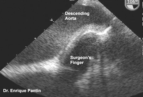
The image below shows the surgeon's finger (in the center) pushing into the aorta of another patient. This aorta was deaired using a cannula in the proximal ascending portion approximately four centimeters above the aortic valve. It is not possible to image this area of the aorta with transesophageal echocardiography due to the interposition of the trachea between the esophagus and that portion of the aorta.

Back to E-chocardiography Home Page.

The contents and links on this page were last verified on
October 24, 2012
by Dr. Olga Shindler.