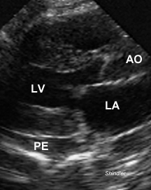
Increased left ventricular wall thickness, abnormal myocardial reflectivity and a small pericardial effusion. This combination of findings suggests amyloid infiltrative cardiomyopathy.
Eur J Echocardiogr. 2006 Jan;7(1):22-30.
Pulsed tissue Doppler and strain imaging discloses early signs of infiltrative cardiac disease: a study on patients with familial amyloidotic polyneuropathy.
Lindqvist P, Olofsson BO, Backman C, Suhr O, Waldenstrom A.
Department of Public Health and Clinical Medicine, Heart Centre, Umea University, S-90185 Umea, Sweden. per.lindqvist@medicin.umu.se
BACKGROUND: Familial amyloidotic polyneuropathy (FAP) is a hereditary systemic amyloidosis with cardiac involvement. As early identification of the cardiac involvement is of major clinical interest we performed this study to test the hypothesis that tissue Doppler imaging (TDI) and strain imaging (SI) might disclose cardiac involvement in patients with early stages of FAP. METHODS: Twenty-two patients with FAP and 36 healthy controls were studied. Standard M-mode and Doppler echocardiography were performed. TDI and SI were used to assess the regional longitudinal left ventricular (LV) lateral and septal and right ventricular (RV) wall functions. All time intervals were corrected for heart rate by dividing with R-R interval and presented as percentage. RESULTS: We found that patients in comparison with controls had increased LV and RV wall thickness and by using TDI a prolonged isovolumic relaxation time (IVRt) at the septal segment (15.0+/-7.0 vs 10.7+/-4.1%, p<0.05) and prolonged isovolumic contraction time (IVCt) at LV lateral (12.8+/-4.3 vs 10.1+/-3.3%, p<0.05), septal (12.5+/-3.5 vs 8.9+/-1.9%, p<0.001) and RV free wall segments (12.0+/-3.6 vs 8.3+/-2.1%, p<0.001). Strain was reduced at LV lateral basal segment (-4.6+/-14.0 vs -20.2+9.1, p<0.001), RV free wall mid segment (-16.2+/-12.8 vs -29.4+/-15.2) as well as both septal segments (-4.1+/-11.7 vs -16.2+/-9.0%, p<0.001, -8.8+/-11.5 vs - 19.4+/-8.4%, p<0.001 for septal basal and mid-segment). Even in the absence of septal hypertrophy the septal strain was reduced and the regional IVCt was prolonged. CONCLUSIONS: This is the first clinical study using TDI and strain in patients with FAP showing functional abnormalities before any morphological echocardiographic abnormalities were present. Both the left and right heart functions are involved and the disease should therefore be regarded as biventricular.
Eur Heart J. 2005 Jan;26(2):173-9. Epub 2004 Dec 9.
Left atrial myopathy in cardiac amyloidosis: implications of novel echocardiographic techniques.
Modesto KM, Dispenzieri A, Cauduro SA, Lacy M, Khandheria BK, Pellikka PA, Belohlavek M, Seward JB, Kyle R, Tajik AJ, Gertz M, Abraham TP.
Division of Cardiovascular Diseases, Mayo Clinic College of Medicine, Rochester, MN, USA.
AIMS: To assess left atrial (LA) function and determine the prevalence of LA dysfunction in AL amyloidosis (AL) using conventional and strain echocardiography. METHODS AND RESULTS: LA ejection fraction, LA filling fraction, LA ejection force, peak LA systolic strain rate (LAsSR), and LA systolic strain (LA epsilon) were determined in 95 AL patients (70 with and 25 without echocardiographic evidence of cardiac involvement, abbreviated CAL and NCAL, respectively), 30 age-matched controls (CON), and 20 patients with diastolic dysfunction and LA dilatation (DD). Peak LAsSR >2 standard deviations below mean CON value was used as the cut-off for normal LA function. LA ejection fraction was lower in CAL when compared with CON (40.4+/-13.6 vs. 67.0+/-6%, P=0.01). Left atrial septal strain rate and strain were lower in CAL (0.8+/-0.5 s(-1) and 5.5+/-4%, respectively) compared with CON (1.8+/-0.8 s(-1) and 14+/-4%, respectively, P=<0.0001), NCAL (1.6+/-0.8 s(-1) and 13+/-7%, respectively, P<0.0001) and DD (1.3+/-0.4 s(-1) and 10+/-2%, respectively, P<0.0001). Based on peak LA systolic strain rate criteria, the cut-off values for normal LA function were -1.1 s(-1) and -1.05 s(-1) for lateral and septal walls. Using these criteria, LA dysfunction was identified in 32% (lateral LA criteria) and 60% (septal LA criteria) of CAL patients. Lateral and septal LAsSR were lower in CAL patients with vs. those without symptoms of heart failure. Inter- and intra-observer agreement was high for LA strain echocardiography. CONCLUSION: LA function assessment using strain echocardiography is feasible with low intra- and inter-observer variability. LA dysfunction is observed in AL patients without other echocardiographic features of cardiac involvement and may contribute to cardiac symptoms in CAL.
Ren Fail. 2005;27(4):415-20
Tissue Doppler is a more reliable method in early detection of cardiac dysfunction in patients with AA amyloidosis.
Demir M, Paydas S, Cayli M, Akpinar O, Balal M, Acarturk E.
Department of Cardiology, School of Medicine, Qukurova University, Adana, Turkey.
OBJECTIVE: Cardiac deposition of AA amyloidosis may result in increasing left ventricular mass and systolic and diastolic dysfunction (DD). The aim of this study was to investigate the left ventricular systolic and diastolic functions by both tissue Doppler imaging (TDI) and pulsed wave Doppler echocardiography (PWD) in patients with AA amyloidosis without congestive heart failure symptoms or arrthymia. METHODS AND RESULTS: Twenty-four patients with AA amyloidosis without congestive heart failure symptoms or arrthymia (15 men and nine women; mean age 44.3 +/- 16.7 years) and 25 healthy subjects (19 men and six women; mean age 43.1 +/- 9.2 years) as controls were included in the study. M-mode, two-dimensional, PWD, and TDI were performed. Peak transmitral filling velocity (E wave), peak transmitral atrial filling velocity (A wave), deceleration time, and isovolumic relaxation time were measured by PWD recordings. Peak myocardial systolic velocity (Sm), peak myocardial early (Em), and late diastolic velocities (Am) were also recorded by TDI. E/A ratio less than one was accepted as DD for both methods. Ejection fraction (EF) was calculated by Teicholtz method. The subjects were divided into three groups as follows: healthy controls (group 1), patients without DD (group 2), and patients with DD (group 3) according to the PWD findings. PWD echocardiography showed that DD was present in 50% of the patients, whereas TDI showed DD in 66% of such cases. In subgroup analysis, Sm wave as a systolic function index was lower in group 3 than in groups 1 and 2, whereas mean EF values were similar in all groups. CONCLUSION: Although AA amyloidosis uncommonly causes cardiac symptoms and findings, according to our results, patients with AA amyloidosis may have systolic and diastolic dysfunction eventhough they are asymptomatic. Also, tissue Doppler imaging is a more reliable method in the early detection of cardiac dysfunction in such patients.
Med Arh. 2005;59(6):388-90.
Echocardiographic assessment of diagnosis and prognosis of biopsy-proven amyloid cardiomyopathy
Sokol I, Vincelj J, Saric M.
Zavod Za Kardiovaskularne Bolesti, Klinika Za Unutarnje Bolesti Klinicka Bolnica Dubrava, Zagreb, Hrvatska. ISOKOL@KBD.HR
Amyloid cardiomyopathy is myocardial infiltrative disorder which mostly has been seen as the consequence of systemic amiloidosis. The diffuse global myocardial infiltration of nonfunctional amyloid displaces the contractile myocites giving rice to relaxation abnormality and diastolic dysfunction of restrictive or congestive type of both ventricles, but more frequently with right-sided congestion, while systolic left ventricular function deteriorates late in disease process. We report a patient with amyloid cardiomyopathy and nephrotic syndrome underlying primary amiloidosis. Our aim is to point out at echocardiographic assessment of diagnosis and prognosis of amyloid cardiomyopathy, which is proven by postmortal endomyocardial biopsy. The hallmark of echocardiographic diagnostics are the findings of the thickened ventricular and septal walls, small ventricular cavities, dilated atria with thickened interatrial septum and atrioventricular valves, and granular-sparkling and hyperrefractile myocardium. Doppler assessment diagnostically gives us the insight in restrictive physiology of both ventricles, and the inverse relation of the left ventricular thickness and voltage on the ECG is high specific. Echocardiographic evaluation of mean left ventricular thickness in amyloid cardiomyopathy is very important prognostic parameter. so that if it is > or =15 mm, median survival is 0.4 years, whereas in our patient with median thickness of 2.76 cm the survival was only three months. The advanced diastolic dysfunction of the left ventricle with an increased transmitral E/A ratio and deceleration time of < or =150 ms is strong predictor of cardiac death. In this case of restrictive transmitral flow E/A was 1.7 and DT 100 ms and they were ominous prognostic signs of survival.
Adv Ther. 2005 Sep-Oct;22(5):433-42.
Doppler tissue imaging of the heart in secondary amyloidosis.
Ulucam M, Yildirir A, Muderrisoglu H, Sezer S, Ozdemir N.
Cardiology Department, Baskent University, Ankara, Turkey.
Secondary amyloidosis (SA) affects cardiac texture and function by interstitial fibrosis. Doppler tissue imaging (DTI) may quantify heart function through the assessment of myocardial velocities. Echocardiographic findings of early cardiac amyloidosis (CA) without heart failure (HF) caused by SA were determined both by standard methods and DTI. It was then determined whether DTI is superior to conventional echocardiography in documenting early CA due to SA. Twenty-five patients with SA who had CA without HF (group 1) were compared with 25 healthy control subjects (group 2). After standard echocardiography, systolic (s), early (e) and late diastolic (a) velocities of interventricular septum, anterolateral, and anterior and inferior walls were measured from mitral annulus by DTI. The averages were called (s(mean)), (e(mean)), and (a(mean)), respectively. Fractional shortening (FS) and ejection fraction (EF) values of groups 1 and 2 were similar. Standard Doppler echocardiographic values were not typical for a specific diastolic abnormality. The (s(mean)) and (e(mean)) for group 1 were lower but (a(mean)) was higher compared with group 2 (all P<.05). The group 1 (e(mean)/a(mean)) was lower (P<.0001) and (E/e(mean)) was higher (P=.003) than in group 2 (both P<.05). (E/e(mean)) and (E/e(lateral wall)) ratios were positively correlated (r=0.74, P<.05). In patients with early CA due to SA without HF, by DTI, (s(mean)) and (e(mean)) velocities decrease and (a(mean)) velocity increases. These may be markers of subclinical CA of SA when standard echocardiography is not informative. (E/e(mean)) ratio may be an alternative index to (E/e(lateral wall)).
Amyloid. 2005 Dec;12(4):246-50.
Regionally heterogeneous tissue mechanics in cardiac amyloidosis.
Petre RE, Quaile MP, Wendt K, Houser SR, Wald J, Goldman BI, Margulies KB.
Cardiovascular Research Center, Temple University School of Medicine, Philadelphia, PA, USA.
OBJECTIVE: The goal of this study was to examine in vitro tissue stiffness and contractile performance in myocardial amyloidosis.BACKGROUND: Primary systemic amyloidosis involves the deposition of amyloid protein in mesodermal tissues including the heart. Functional assessment of cardiac amyloidosis is usually performed using echocardiography. However, this technique does not involve assessment of preload-dependent contractile reserve (the Frank-Starling mechanism).METHODS: At the time of heart transplantation, isolated myocardial trabeculae were dissected from the right ventricle of a patient with primary systemic amyloidosis. In vitro length-tension experiments were performed and trabeculae were subsequently fixed, sectioned and stained with crystal violet to determine amyloid deposition.RESULTS: Among the nine trabeculae capable of generating force transients, various combinations of myocardial stiffness and contractile performance were observed including normal stiffness and contractility, severely increased stiffness with impaired contractility and hybrid patterns. Histological analysis demonstrated varying degrees of amyloid deposition among sampled trabeculae.CONCLUSIONS: Our findings extend previous reports of functional heterogeneity among patients by demonstrating functional heterogeneity within a single patient's heart. Our findings also highlight the functional interdependence of passive stiffness and systolic performance in the diseased myocardium and demonstrate the value of dynamic assessments of myocardial performance.
Cardiovasc J S Afr. 2004 May-Jun;15(3):136-8.
Cardiac amyloidosis presenting as pseudo-hypertrophic cardiomyopathy.
Papachan A, Sliwa K, Gildenhuys A, Essop R.
Department of Cardiology, Chris Hani-Baragwanath Hospital, Johannesburg, South Africa.
Both cardiac amyloidosis and hypertrophic cardiomyopathy may result in excessive hypertrophy of the myocardium, which can be seen on echocardiography. While in most patients the two conditions are easily differentiated, we present in this report a case of amyloidosis that mimicked hypertrophic cardiomyopathy so closely that it required endomyocardial biopsy to establish the diagnosis.
Curr Opin Cardiol. 2004 Sep;19(5):464-71.
Tissue Doppler imaging in the evaluation of patients with cardiac amyloidosis.
Sallach JA, Klein AL.
Section of Cardiovascular Imaging, Department of Cardiovascular Medicine, The Cleveland Clinic Foundation, Cleveland, Ohio 44195, USA.
PURPOSE OF REVIEW: Although two-dimensional, M-mode, and Doppler echocardiography have played a major role in the assessment of amyloid deposition in the heart, diagnosis of cardiac amyloidosis (CA) based on these conventional techniques is often only possible once the disease is in a relatively advanced stage. To optimize survival, early diagnosis and institution of therapy are essential. Recently, tissue Doppler imaging (TDI) and myocardial strain rate (SR) have emerged as important clinical tools in the assessment of CA. RECENT FINDINGS: Tissue Doppler imaging-derived modalities including TDI velocities, strain, and SR are currently being used in the early diagnosis and evaluation of patients with CA. Although these new indices have been examined in relatively few patients, findings suggest an important and expanding role of TDI in amyloid infiltration of the heart. SUMMARY: This review summarizes the recent literature addressing the role of TDI velocities, strain, and SR in the diagnosis and assessment of CA.
American Journal of Transplantation Volume 1 Page 93 - April 2001 doi:10.1034/j.1600-6143.2001.010117.x Volume 1 Issue 1
Fatal Disseminated Aspergillosis following Sequential Heart and Stem Cell Transplantation for Systemic Amyloidosis Raymund R. Razonablea, Robin Patela,b,*, Mark P. Wilhelma, Morie A. Gertzc, Mark R. Litzowc, David J. Inwardsc, Joseph A. Dearanid, Brooks S. Edwards and Christopher G. McGregord
Infectious complications are a major cause of morbidity and mortality in transplant recipients. We describe a case of fatal disseminated aspergillosis immediately following autologous peripheral stem cell reconstitution in a patient who had undergone orthotopic heart transplantation for systemic amyloidosis. The case described suggests that the infectious risks in patients undergoing these sequential procedures may be distinct from those occurring in patients undergoing either procedure independently. Potential prophylactic and therapeutic interventions are discussed. Since this experimental and evolving approach for the management of systemic amyloidosis is potentially applicable to a limited number of patients, multicenter collaboration may be needed to further define the infectious risks in this unique subset of transplant recipients.
Arch Mal Coeur Vaiss. 1993 Jul;86(7):1009-15.
Technetium TC 99m pyrophosphate myocardial scintigraphy in amyloidosis. Correlations with Doppler echocardiography
Fournier C, Grimon G, Rinaldi JP, Terral A, Boujon B, Adams A, Desgrez A, Blondeau M.
Service de cardiologie, CHU Bicetre, Le Kremlin-Bicetre.
Technetium 99m cardiac scintigraphy as practiced at present for diagnosing amyloisodid only provides a visual semi-quantitative assessment of uptake of the isotope. To improve the diagnostic accuracy of the method, the authors evaluated prospectively a personal technique of scintigraphy quantification based on early images obtained at the 20th minute in 15 patients with neuropathic amyloidosis. Doppler echocardiographic studies indicated that 9 patients had cardiac involvement whilst 6 were free of cardiac amyloidosis. The index of isotopic uptake (ratio of cardiac/abdominal uptake) was 0.44 to 1.58 in the first group and 0.09 to 0.31 in the second group. The correlation between the scintigraphic index and interventricular septal or posterior wall thickness measured by echocardiography was poor. These results obtained in 15 patients with neuropathic amyloidosis suggest that the scintigraphic index measured at the 20th minute is discriminatory and allows identification of those patients with cardiac involvement. On the other hand, the correlations with echocardiographic wall thickness are poor. Technetium 99m cardiac scintigraphy with this technique of quantification is a useful tool for diagnosing cardiac amyloidosis, especially when echocardiography is difficult to interpret.
Echocardiography. 1991 Mar;8(2):253-9.
Two-dimensional echocardiography in myocardial amyloidosis.
Picano E, Pinamonti B, Ferdeghini EM, Landini L, Slavich G, Orlandini A, Marini C, Lattanzi F, Camerini F.
C.N.R. Clinical Physiology Institute of Pisa, Trieste, Italy.
Two-dimensional echocardiography is the best means of identifying early cardiac amyloid infiltration and gauging its subsequent progression. The early asymptomatic phase is characterized on echocardiography by a mild-to-moderate increase in left ventricular and/or right ventricular wall thicknesses. The distinctive combination of low electrocardiography voltage and increase in left ventricular mass on the echocardiogram, both compatible with substantial amyloid infiltration, is valuable in diagnosis and appears to indicate the severity of the disease. Other ancillary but common findings are left atrial dilatation, a small pericardial effusion, thickening of cardiac valves, papillary muscles, and interatrial septum. Finally, there is a peculiar texture of myocardial walls, with highly refractile areas that are typical, although not specific, of myocardial amyloidosis and can also be quantitatively described by digital image analysis techniques. The echocardiographic appearance of amyloidosis can closely mimic several other diseases. Asymmetric hypertrophy of the septum due to amyloid deposition may occur, simulating hypertrophic cardiomyopathy. The granular sparkling of myocardial walls is also found in myocarditis with severe fibrosis, and it is quite common in hypertrophic cardiomyopathy, as well as in other infiltrative diseases of the myocardium. It is not uncommon that the echocardiographic examination represents a turning point in the work-up of the patient, briskly orienting the clinician towards the correct diagnostic pathway. However, the likelihood of the cardiologist-echocardiographer to successfully and prospectively identify myocardial amyloidosis is substantially higher if all the clinical and electrocardiographic information is reviewed at the time of the echocardiographic examination.
Arch Mal Coeur Vaiss. 1984 Dec;77(13):1525-31.
M-mode and two-dimensional echocardiography of 7 cases of cardiac amyloidosis
Roudaut R, Haissaguerre M, Dallocchio M.
Although rare, cardiac amyloidosis is the commonest cause of infiltrative myocardiopathy. The diagnosis may be suspected clinically in patientswith mainly right ventricular failure of sudden onset. The aim of this study was to assess the diagnostic value of M-Mode and 2D echocardiography in this condition. Seven cases of cardiac amyloidosis were studied. Biventricular hypertrophy, usually more severe on the left side with reduction in size of the left ventricular chamber, was observed in all cases. Parameters of systolic and diastolic function were abnormal. A significant pericardial effusion was demonstrated in 3 patients. 2D echocardiography also allows evaluation of the myocardial structure: in 3 cases the whole of the left ventricular myocardium seemed granular, sparkling and abnormally echogenic. In patients with cardiac failure these appearances are very suggestive of amyloidosis, especially when the ECG shows low voltage complexes and pathological Q waves. In 3 other patients, this abnormal echogenic myocardial appearance was observed only in the interventricular septum, which is much less suggestive of cardiac amyloidosis. In conclusion, in patients with cardiac failure with cardiomegaly and a low voltage ECG, echocardiographic findings of hypertrophic cardiomyopathy (only rarely with dilatation) and hypokinetic wall motion are suggestive of cardiac amyloidosis, especially when the myocardium has a granular, sparkling appearance.
Back to E-chocardiography Home Page.

The contents and links on this page were last verified on
October 24, 2012
by Dr Olga Shindler.