Cardiac Anatomy
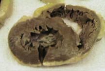 Short axis view of the adult heart illustrating the circular and more
muscular left ventricle, the crescent shaped and more trabeculated
right ventricle. The yellow epicardial fat may appear as an echo free
space on 2D and M-mode echocardiographic images.
Abnormal adiposity of the right heart can be clinically significant.
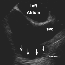 The right atrial appendage is broad based and can be difficult to identify
during transesophageal echocardiography. It can be distinguished from
the smooth right atrial surface by the presence of pectinate muscles
(arrows) which appear as an irregular, lumpy surface.
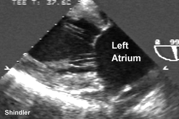
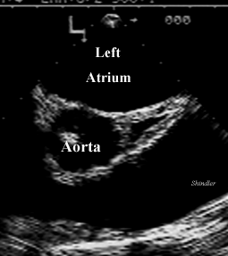 The transverse sinus is a pericardial space that can be confused
with a coronary artery during transesophageal echocardiography. There
is no color flow and it has a characteristic triangular appearance.
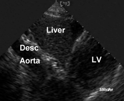 The subcostal view illustrates the relationship of the heart to
the liver and to the aorta.
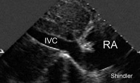 Eustachian valve.
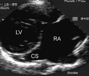 Coronary sinus.
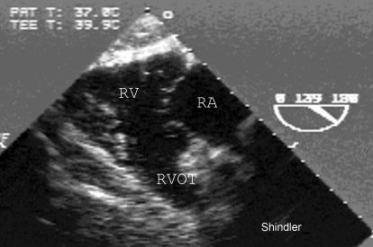 Right ventricle.
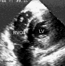 Right ventricular outflow, or conus, or crista supraventricularis,
or superior limbic band.
 Chordal attachments to the mitral valve.
The Azygos and Hemiazygos Veins:
The azygos vein returns blood from the suprarenal segment
of the inferior vena cava to the right superior vena cava.
It passes through the aortic opening of the diaphragm and
courses medial to the thoracic vertebrae. It turns anteriorly
at the level of the fourth thoracic vetebra connecting to
the posterior aspect of the superior vena cava.
The hemiazygos takes off from a lumbar or renal vein passing
through the left crus of the diaphragm, ascends to the left
of the spine turning right at the ninth thoracic vertebral level
behind the thoracic duct and aorta, draining into the azygos vein.
There is also a left upper hemiazygos.
Back to E-chocardiography Home Page.

The contents and links on this page were last verified on
October 24, 2012
by Dr. Olga Shindler.
 Short axis view of the adult heart illustrating the circular and more
muscular left ventricle, the crescent shaped and more trabeculated
right ventricle. The yellow epicardial fat may appear as an echo free
space on 2D and M-mode echocardiographic images.
Abnormal adiposity of the right heart can be clinically significant.
 The right atrial appendage is broad based and can be difficult to identify
during transesophageal echocardiography. It can be distinguished from
the smooth right atrial surface by the presence of pectinate muscles
(arrows) which appear as an irregular, lumpy surface.
 The transverse sinus is a pericardial space that can be confused
with a coronary artery during transesophageal echocardiography. There
is no color flow and it has a characteristic triangular appearance.
 The subcostal view illustrates the relationship of the heart to
the liver and to the aorta.
 Eustachian valve.
 Coronary sinus.
 Right ventricle.
 Right ventricular outflow, or conus, or crista supraventricularis,
or superior limbic band.
 Chordal attachments to the mitral valve.
