Chronic Descending Aortic Dissection
by: Paul Haluska, M.D.
-
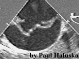
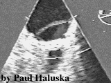
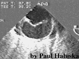 Spontaneous contrast in the
false lumen and entry tear.
Spontaneous contrast in the
false lumen and entry tear.
-
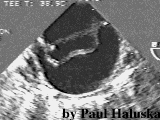
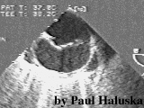 Sequential tomographic cuts
obtained by advancing the transesophageal echo probe.
Sequential tomographic cuts
obtained by advancing the transesophageal echo probe.
-
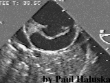
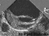 Partial thrombosis of the false lumen.
-
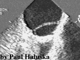
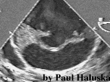 Grayscale flow Doppler in the true lumen
and in the entry tear.
Grayscale flow Doppler in the true lumen
and in the entry tear.
-
 Less thrombus in the false lumen a few
millimeters lower.
Less thrombus in the false lumen a few
millimeters lower.
-
 Cyclical compression of the true lumen.
-
 Lung parenchyma is shown in addition to the dissection.
-
 Small caliber of true lumen compared
to the false lumen at the gastroesophageal juntion.
Small caliber of true lumen compared
to the false lumen at the gastroesophageal juntion.
-
 Grayscale flow in the true lumen.
Back to E-chocardiography Home Page.
e-mail:shindler@umdnj.edu