Echocardiographic features in the neonate:
Dilated right cardiac chambers, small left cardiac chambers, right-to-left displacement of the interatrial septum.
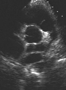
The abnormal confluence chamber behind the left atrium may be small and difficult to visualize when pulmonary vein flow is decreased.
Auscultation in the neonate:
Increased flow across the pulmonic valve may give rise to a systolic murmur.
Increased flow across the tricuspid valve may give rise to a diastolic murmur.
Auscultation in the neonate may not reveal any murmurs if pulmonary blood flow is decreased due to elevated pulmonary vascular resistance.
The second heart sound is split in this cyanotic lesion. Other cyanotic lesions in the neonate such as transposition or pulmonary atresia have a single second sound.
Int J Cardiol. 2005 Nov 9;
Total anomalous pulmonary venous return in the fourth decade.
Nurkalem Z, Gorgulu S, Eren M, Bilal MS.
Cardiology Department, Siyami Ersek Thoracic and Cardiovascular Surgery Center, Istanbul, Turkey.
Most patients with total abnormal venous connection (TAPVC) have no symptoms at birth, yet the majority die within the first year of life if surgical repair is not implemented. TAPVC is usually suspected in the neonatal period, and without surgical repair prolonged survival is quite exceptional. This kind of abnormality can be a real challenge for a non-pediatrics cardiologist today and can be easily misdiagnosed as a large secundum atrial septal defect if there is no sufficient suspicion of TAPVC. Therefore, a case of a 40-year-old female patient referred to our clinic with increasing shortness of breath is of great interest, as this patient was diagnosed with TAPVC.
Ann Thorac Surg. 2005 Sep;80(3):1140-2.
Modified septosuperior approach for the repair of supracardiac total anomalous pulmonary venous return in infants.
Balakrishnan KR, Parvathy U.
Department of Cardiothoracic Surgery, Sri Ramachandra Medical College University Hospital, Chennai, India. krbalakrishnan@vsnl.com
An alternative technique for the repair of supracardiac total anomalous pulmonary venous return (TAPVR) is described. The pulmonary venous confluence (CPV) is identified below the right pulmonary artery between the aorta and superior vena cava. The atrial incisions are similar to a septosuperior approach of the mitral valve, modifying the incision on the roof of the left atrium to be parallel to the incision in the CPV. The common pulmonary vein is anastomosed to the roof of the left atrium. This approach offers optimal exposure for the repair without distorting the cardiac structures.
J Thorac Cardiovasc Surg. 2005 May;129(5):1091-7.
Late neurodevelopmental outcome after repair of total anomalous pulmonary venous connection.
Kirshbom PM, Flynn TB, Clancy RR, Ittenbach RF, Hartman DM, Paridon SM, Wernovsky G, Spray TL, Gaynor JW.
Division of Cardiovascular Surgery, Emory University, Emory Cklinic Building A, Atlanta, GA 30322, USA. paul_kirshbom@emoryhealthcare.org
Objective We sought to define the neurodevelopmental status of school-aged survivors of total anomalous pulmonary venous connection repaired during infancy. Methods All school-aged survivors of total anomalous pulmonary venous connection repair performed at a single institution were eligible. Thirty children returned for neurologic examination and neurodevelopmental testing. Results The median age at total anomalous pulmonary venous connection repair was 16 days (range, 1-141 days), and age at testing was 11 years (range, 6-19 years). Pulmonary venous return was supracardiac in 14 patients, infracardiac in 12 patients, cardiac in 3 patients, and mixed in 1 patient. Preoperative obstructed total anomalous pulmonary venous connection was present in 6 patients. Circulatory arrest was used in all repairs, with a median duration of 35 minutes (range, 17-55 minutes). At follow-up, microcephaly (head circumference <5%) was present in 28%. Neuromuscular examination was suspect or abnormal in 27%. Mean Full-scale IQ (95.3 +/- 18.5) and Verbal IQ (98.6 +/- 20.2) were not different from population norms, but Performance IQ (92.3 +/- 16.9) was significantly lower than population norms ( P = .02). Fine motor skills and visual-motor coordination were significantly impaired ( P < .01 for Grooved Pegboard and Test of Visual-Motor Integration). Patients with total anomalous pulmonary venous connection also had difficulty with tests of attention (Test of Everyday Attention for Children, P < .01), but results of tests of memory function were not significantly different from population norms. Conclusions School-aged survivors of infant total anomalous pulmonary venous connection repair exhibit a significant incidence of neurodevelopmental difficulties. Fine motor function, visual-motor integration, and attention are the most commonly affected domains. Evaluation of these children is indicated to identify those who are at risk for learning disabilities and who could benefit from early intervention.
Ann Thorac Surg. 2004 Dec;78(6):2186.
Three-dimensional demonstration of total anomalous pulmonary venous return with contrast-enhanced magnetic resonance angiography.
Yoshioka K, Niinuma H, Kawakami T, Oyama K, Ishihara K, Kawazoe K.
Department of Radiology,Memorial Heart Center, Iwate Medical University, Morioka, Japan. kyoshi@iwate-med.ac.jp
A newborn male was hospitalized for a heart murmur and severe cyanosis after his birth. Echocardiography showed an abnormal vein crossing below the diaphragm; total anomalous pulmonary venous return (TAPVR, Darling type III) was suspected. A contrast-enhanced 3-dimensional magnetic resonance angiography (MRA) was performed on the 6th day after his birth to assess pulmonary vein stenosis. The MRA was obtained with a 1.5-T superconducting imager (Signa Horizon LX EchoSpeed, with 8.3 operating system software; GE Medical Systems, Milwaukee, WI) and a 4-channel phased array coil for the brain to optimize signal detection with the image sequence from a fast-spoiled gradient-echo (fast-SPGR) with fat suppression. Contrast medium (gadopentetate dimeglumine, 0.1 mmol/kg, Magnevist, Schering, Berlin, Germany), 0.6 mL, was administered at 0.2 mL/sec using a power injector.
J Chin Med Assoc. 2004 Jul;67(7):331-5.
Significance of pulmonary venous obstruction in total anomalous pulmonary venous return.
Wang PY, Hwang BT, Lu JH, Lee PC, Tiu CM, Weng ZC, Meng LC.
I-Lan Hospital, Department of Health, Taipei, Taiwan, ROC.
BACKGROUND: Total anomalous pulmonary venous return (TAPVR) is an uncommon congenital cardiovascular anomaly with poor natural prognosis. It has been detected more frequently in recent year due to the advent of echocardiography and cardiovascular magnetic resonance imaging (MRI). The aim of this study was to evaluate the clinical manifestations and outcomes in TAPVR patients with or without pulmonary venous obstruction (PVO). METHODS: From January 1985 to December 2002, a total of 27 cases with TAPVR at our institution were reviewed. Accurding to the preseace or assence of PVO, patients were divided into PVO group and non-PVO group. Patients' sex, age at diagnosis, types of TAPVR, clinical manifestations, surgical treatment and outcomes were evaluated. RESULTS: All of them had received 2-dimensional (2-D) and color Doppler echocardiography examination. Cardiac catheterization was performed in all but 1 patient who died at the first day of birth. In addition, 10 of 27 cases had cardiovascular MRI for further study. The number of cases in PVO group and non-PVO group were 15 (56%) and 12 (44%), respectively. There was no significant difference in sex or pulmonary venous drainage sites between both groups. Cyanosis was more prevalent in the PVO group (80% vs. 30%, p = 0.038). Four (27%) cases PVO group and 3 (25%) cases of the non-PVO group had of the non-isolated cardiac lesions. Pulmonary hypertension was present in 18 (69%) of 26 cases who had received cardiac catheterization. Among them, 10 had PVO and 5 had systemic level of pulmonary arterial pressure. Seven (30%) of 23 patients who had received operation died; in contrast, 3 of 4 patients without operation expired. The remaining 1 did not had surgery because of complex heart disease. There was no significant difference in surgical mortality between PVO and non-PVO groups (33% vs. 27%). CONCLUSIONS: Cyanosis is an obvious clinical symptom of obstructed TAPVR. Surgical mortality made no significant difference between obstructed and non-obstructed groups. Early detection and surgical treatment for TAPVR are important. Although cardiac catheterization and angiocardiography is the golden standard for the diagnosis, 2-D and color Doppler echocardiography can also provide quick and accurate diagnostic images of TAPVR.
Turk J Pediatr. 2004 Apr-Jun;46(2):179-81.
Acute pulmonary edema in a newborn with infracardiac type total anomalous pulmonary venous return and surgical repair.
Yalcinbas YK, Erek E, Salihoglu E, Ozturk N, Mamur G, Soybir N, Sarioglu A, Sarioglu T.
Istanbul Memorial Hospital, Istanbul, Turkey.
Total anomalous pulmonary venous return (TAPVR) is a rare congenital pathology. Early diagnosis and urgent surgery are life-saving, especially in newborns with pulmonary venous obstruction, which is most commonly seen with infracardiac type. A three-day-old baby boy presented to another clinic with tachypnea and cyanosis. Initial work-up aimed at ruling out persistant pulmonary hypertension, respiratory distress syndrome and pneumonia. Acute pulmonary edema then developed, and on echocardiography obstructive type infracardiac TAPVR was suspected. Cardiac catheterization was done for definitive diagnosis. Urgent surgery was undertaken and pulmonary veins were anastomozed to left atrium with posterior approach. Patient was extubated at 10th day and discharged after three weeks. During one-year follow-up the patient was free of symptoms. Infracardiac type TAPVR is a rare pathology in which early diagnosis and urgent surgery with special postoperative case are mandatory for survival.
Radiographics. 2004 May-Jun;24(3):755-72.
Erratum in:
Radiographics. 2004 Sep-Oct;24(5):1514.
Congenital hepatic shunts.
Gallego C, Miralles M, Marin C, Muyor P, Gonzalez G, Garcia-Hidalgo E.
Department of Radiology, Hospital Universitario 12 de Octubre, Carretera de Andalucia km 5,400, 28041 Madrid, Spain. mamengallego@terra.es
Abnormal vascular connections within the hepatic parenchyma are occasionally seen at ultrasonography (US) and require further evaluation. The radiologic findings in 42 children with infantile hepatic hemangioma (n = 28), vascular malformations (n = 10), or infradiaphragmatic total anomalous pulmonary venous return (TAPVR) (n = 4) associated with congenital vascular shunting were retrospectively reviewed. Arteriovenous connections are seen in infantile hepatic hemangiomas and arteriovenous malformations and manifest with aortic tapering at the level of the celiac trunk, hepatic artery enlargement with a low resistivity index (RI), and increased flow velocities in the hepatic veins that may assume an arterialized spectral pattern in late-stage disease. Congenital arterioportal shunts demonstrate a low RI in the hepatic artery, hepatofugal arterialized flow in the portal vein, and rapid development of signs of portal hypertension. Portosystemic shunting may be intra- or extrahepatic. A pulsatile triphasic spectral pattern is seen in the portomesenteric venous system in children with portosystemic shunting, and macroscopic connections between the portal system and the hepatic veins are evident. Infradiaphragmatic TAPVR is associated with a tortuous vessel that parallels the aorta, ends at the intrahepatic left portal vein or the ductus venosus, and has hepatopetal flow. Familiarity with the US features of various congenital abnormal hepatic vascular connections will aid in diagnosis and treatment. Copyright RSNA, 2004
Birth Defects Res A Clin Mol Teratol. 2004 Apr;70(4):185-93.
Parental lead exposure and total anomalous pulmonary venous return.
Jackson LW, Correa-Villasenor A, Lees PS, Dominici F, Stewart PA, Breysse PN, Matanoski G.
National Institute of Child Health and Human Development, Rockville, Maryland, USA. jacksole@mail.nih.gov
BACKGROUND: Investigators from the Baltimore-Washington Infant Study (BWIS) reported an association between self-reported maternal lead exposure and total anomalous pulmonary venous return (TAPVR) in their offspring. This association was further evaluated in the BWIS population using a more sensitive exposure estimate. METHODS: Cases included 54 live-born infants with TAPVR; controls were a stratified random sample of 522 live-born infants from the BWIS control group. Parental lead exposure was based on three assessment methods, including: an industrial hygiene assessment, an a priori job exposure matrix, and self-reported exposures. A parent was classified as exposed to lead if he/she was classified as exposed by any one of the assessment methods. RESULTS: Approximately 17% of case mothers and 11% of control mothers were classified as exposed to lead during the three months prior to conception through the first trimester (odds ratio [OR], 1.57; 95% confidence interval [CI], 0.64-3.47). Among fathers, 61% of case fathers and 46% of control fathers were classified as exposed to lead during the six months prior to conception (paternal critical period) (OR, 1.83; 95% CI, 1.00-3.42). During the paternal critical period, when only the father was exposed compared to neither parent exposed, the OR for any lead exposure and TAPVR was 1.65 (95% CI, 0.84-3.25). CONCLUSIONS: This study supports a possible association between paternal lead exposure and TAPVR. Further studies are warranted using validated assessment methods for occupational and nonoccupational lead exposures to corroborate this association and to elucidate the possible biological mechanism. Birth Defects Research (Part A), 2004. Copyright 2004 Wiley-Liss, Inc.
Radiology. 2004 Mar;230(3):824-9. Epub 2004 Jan 22.
Variations in pulmonary venous drainage to the left atrium: implications for radiofrequency ablation.
Marom EM, Herndon JE, Kim YH, McAdams HP.
Department of Radiology, Duke University Medical Center, Durham, NC, USA. emarom@di.mdacc.tmc.edu
PURPOSE: To evaluate and classify the various drainage patterns of the pulmonary veins as depicted with thin-section chest computed tomography (CT). MATERIALS AND METHODS: Thin-section (2.5-mm collimation) contrast material-enhanced CT scans of 201 consecutive patients obtained over a 3-month period for diagnosis of pulmonary embolism (n = 197), pulmonary vein stenosis (n = 2), or aortic injury (n = 2) were routinely reviewed in transverse and (if necessary) coronal and coronal-oblique imaging planes. A classification was formulated based on both the number of venous ostia on each side and the drainage patterns of pulmonary veins. The frequency of each pattern was determined, and association with atrial arrhythmia was assessed with the chi(2) and Fisher exact tests. RESULTS: Most patients (n = 142, 71%) had two ostia on the right side for upper and lower lobe veins. Fifty-six patients (28%) had three to five ostia on the right side, which were due to one or two separate middle lobe vein ostia in 52 (26%) patients. Three patients (2%) had a single venous ostium on the right side. Most patients (n = 173, 86%) had two ostia on the left side for upper and lower lobe veins. The remainder (n = 28, 14%) had a single ostium. There was no significant association between any particular venous drainage pattern and atrial arrhythmia; however, patients with a separate ostia for the right middle lobe pulmonary vein(s) tended to have a higher frequency of atrial arrhythmia than those with other patterns (P =.053). CONCLUSION: A classification system to succinctly describe pulmonary venous drainage patterns was developed. Right-sided venous drainage was more variable than left-sided venous drainage. One-quarter of patients had more than two venous ostia on the right side. Copyright RSNA, 2004
Jpn J Thorac Cardiovasc Surg. 2002 Aug;50(8):338-40.
In situ pericardium repair of pulmonary venous obstruction after repair of total anomalous pulmonary venous connection.
Nishi H, Nishigaki K, Kume Y, Miyamoto K.
Department of Pediatric Cardiovascular Surgery, Osaka City General Hospital, 2-13-22 Miyakojima Hondori, Miyakojima-ku, Osaka 534-0021, Japan.
A 13-month-old boy with recurrent pulmonary venous obstruction (PVO) after repair of total anomalous pulmonary venous connection (TAPVC, Darling IIa + Ia) was treated successfully with in situ pericardium repair consisting of unroofing coronary sinus at 2 months. At 8 months, stenosis of the right upper and lower pulmonary veins (PV) and left lower PV were detected, and PVO was relieved via resection of the stenosis site and recutback. Echocardiography 3 months later showed obstructed bilateral PVs and connection between left PVs and vertical veins. At reoperation, we conducted in situ pericardium repair for right PVO and anastomosed left PVs to the left atrial appendage. The postoperative course was satisfactory. Echocardiography 12 months later showed no evidence of PVO, but cardiac catheterization 12 months later showed mild obstruction on the right side and normal venous drainage on the left. Although the long-term prognosis is unknown, this sutureless technique is effective in recurrent PVO.
Indian Heart J. 1999 Jan-Feb;51(1):65-8. Related Articles, Links
Mixed variety of total anomalous pulmonary venous connection: diagnosis by 2D echocardiography and Doppler colour flow imaging.
Saxena A, Reddy SC, Kothari SS, Juneja R, Venugopal P, Shrivastava S.
Cardiothoracic Centre, All India Institute of Medical Sciences, New Delhi.
Of the many types of total anomalous pulmonary venous connection, mixed type is the least common. Its accurate non-invasive diagnosis by echocardiography poses a diagnostic challenge. We report our experience of echocardiography in 21 infants with mixed type of total anomalous pulmonary venous connection aged 25 days to one and half years. Multiple windows were used to identify individual pulmonary veins and various sites of drainage. Cardiac catheterisation and angiography were performed for 17 cases. In 11 of 21 cases, the left upper pulmonary vein was seen draining into vertical vein and the left lower and right-sided pulmonary veins were draining into the coronary sinus. Cardiac and supracardiac combinations of other types were seen in eight more cases. Both drainage sites were supracardiac in one case and supracardiac and infracardiac in another. On comparing echocardiographic findings with those obtained at cardiac catheterisation and/or surgery (carried out in 18 cases), there were three instances of error. In two cases (echocardiography performed without the use of colour flow imaging) the second site of drainage could not be defined. These patients were catheterised as all four pulmonary veins were not delineated by echo. The third error occurred in a case where although two sites of drainage were picked up by echo but at surgery, the right lower pulmonary vein was noted to have a double connection, both to coronary sinus and to vertical vein. We conclude that mixed type of total anomalous pulmonary venous connection can be accurately diagnosed by echocardiography and Doppler colour flow imaging. The diagnostic errors are rare and would not alter the surgical management.
Vestn Khir Im I I Grek. 1998;157(3):50-2.
Surgical methods for correcting partial anomalous drainage of the pulmonary veins during artificial circulation
Makhmudov MM, Guliamov DS, Mamatov MA.
The authors share their experiences with surgical treatment of 42 patients with different anatomical variants of the partial anomalous drainage of the pulmonary veins. Specific features of the surgical strategy are described depending on the level of confluence of the pulmonary veins, especially if the mouths of the abnormally falling pulmonary veins and the interatrial septum defect are far from each other. Good immediate and nearest results were obtained.
Z Kardiol. 1998 Apr;87(4):288-92.
Echocardiography diagnosis of a partial anomalous pulmonary vein anastomosis in 2 patients with Ullrich-Turner syndrome
Koch A, Hofbeck M, Dorr HG, Singer H.
Klinik mit Poliklinik fur Kinder und Jugendliche, Universitat Erlangen-Nurnberg.
We report on 2 infants with Ullrich-Turner syndrome in whom partial anomalous pulmonary venous drainage was diagnosed noninvasively by color-coded Doppler sonography. Several patients with the combination of anomalous drainage of one or more pulmonary veins and Ullrich-Turner syndrome have been described in the literature. However, in the majority of those previously reported cases the diagnosis of partial anomalous pulmonary venous drainage was established by angiography during cardiac catheterization performed for confirmation of other cardiovascular malformations. Our patients show that partial anomalous pulmonary venous drainage can be diagnosed easily in neonates and young infants, as long as this anomaly is taken into consideration.
Ultrasound Obstet Gynecol. 1997 May;9(5):347-9.
Abnormal pulmonary venous return diagnosed prenatally by pulsed Doppler flow imaging.
Feller Printz B, Allan LD.
Babies' Hospital, Columbia-Presbyterian Medical Center, New York, NY, USA.
Pulsed Doppler ultrasound has been used to characterize distinctive pulmonary venous flow patterns in the normal fetus and child. Changes in these characteristic flow patterns have been related to abnormal atrial and ventricular hemodynamics. We report a case of total anomalous pulmonary venous return diagnosed prenatally because of an abnormal pulsed Doppler echocardiographic flow pattern, even though color flow mapping appeared to demonstrate normal pulmonary venous drainage. This case demonstrates the importance of obtaining pulsed Doppler pulmonary venous flow profiles during fetal echocardiography, especially in cases of complex congenital heart disease.
J Am Coll Cardiol. 1997 May;29(6):1351-8.
Partial anomalous pulmonary venous connection: diagnosis by transesophageal echocardiography.
Ammash NM, Seward JB, Warnes CA, Connolly HM, O'Leary PW, Danielson GK.
Division of Cardiovascular Diseases and Internal Medicine, Mayo Clinic, Rochester, Minnesota 55905, USA. ammash.naser@mayo.edu
OBJECTIVE: This study sought to demonstrate that with proper technique, identification of the normal and abnormal pulmonary venous connection can be made with confidence using transesophageal echocardiography (TEE). BACKGROUND: Partial anomalous pulmonary venous connection (PAPVC) is an uncommon congenital anomaly whose diagnosis has classically been made using angiography. METHODS: We performed a retrospective review of all patients of all ages with PAPVC diagnosed at the Mayo Clinic who had undergone TEE because of either right ventricular volume overload or suspected intracardiac shunting by transthoracic echocardiography or intraoperatively. RESULTS: A total of 66 PAPVCs were detected in 43 patients (1.5/patient); in 2 additional patients, TEE suggested, but did not diagnose, PAPVCs. Shortness of breath was the most common presenting symptom (42.2%), followed by heart murmur and supraventricular tachycardia. Right-sided anomalous veins were identified in 35 patients (81.4%), left-sided in 7 (16.3%) and bilateral in 1 (2.3%). There was a single anomalous connecting vein in 23 patients (53.5%), two in 18 (41.9%), three in 1 (2.3%) and four in 1 (2.3%). The connecting site was the superior vena cava (SVC) in 39 veins (59.1%), right atrial-SVC junction in 6 (9.1%), right atrium in 8 (12.1%), inferior vena cava in 1 (1.5%) and the coronary sinus in 2 (3.0%). Ten anomalous left pulmonary veins were connected by a vertical vein to the innominate vein (15.1%). Sinus venosus atrial septal defect (ASD) was the most common associated anomaly in 22 patients (49%), followed by ostium secundum ASD in 6 and patent foramen ovale in 4. Fifteen patients had an intact atrial septum. Thirty-one patients (68.8%) underwent surgical repair. PAPVC was confirmed in all patients, including the two whose TEE results were suggestive of PAPVC. All 49 PAPVCs detected by TEE preoperatively were confirmed at the time of operation. CONCLUSIONS: TEE is highly diagnostic for PAPVC and can obviate angiography. Accurate anatomic diagnosis may influence the need for medical and surgical management. TEE should be performed in patients with right ventricular volume overload when the precordial examination is inconclusive.
Arch Mal Coeur Vaiss. 1997 Feb;90(2):295-9.
Acquired pulmonary vein stenosis after surgery of complex partial anomalous pulmonary venous drainage in an adult. Diagnostic value of transesophageal echocardiography
Bouffard P, Bourachot ML, Petit J, Houyel L, Angel CY, Nottin R.
Centre chirurgical Marie-Lannelongue, Le Plessis-Robinson.
Surgical correction of partial anomalous pulmonary venous drainage is difficult and may be complicated by acquired postoperative stenosis at the site of reimplantation of the pulmonary veins in the left atrium. Diagnosis should be made quickly because of the very poor prognosis due to acute pulmonary hypertension. The case described by the authors underlines the value of multiplane transesophageal echocardiography with two-dimensional and Doppler analysis for rapid and accurate diagnosis of this complication.
J Am Soc Echocardiogr. 1996 Mar-Apr;9(2):174-81.
Echocardiographic clues and accuracy in the diagnosis of scimitar syndrome.
Shibuya K, Smallhorn JE, McCrindle BW.
Division of Cardiology, the Hospital of Sick Children, Toronto, Ontario, Canada.
Scimitar syndrome was diagnosed in 27 patients seen between July 1974 and May 1993. All available echocardiograms taken before surgery or death were reviewed. Age at presentation ranged from 1 day to 14 years. Dextrocardia or mesocardia was noted in 70%, atrial septal defect in 70%, and increased right ventricular dimension in 70% of the patients. The ratio of the proximal and distal diameters of the right/left pulmonary arteries were reduced 0.68 +/- 0.17 and 0.66 +/- 0.17, respectively. "Blunting" of the right side of the left atrium was seen in all patients with total anomalous right pulmonary venous drainage and none with partial drainage. Anomalous right pulmonary venous drainage was characterized in 91% of echocardiograms with color flow mapping versus 14% without color flow mapping (p < 0.0002). Aortopulmonary collateral arteries were detected in all four cases in which color flow mapping was performed, but not detected otherwise.
Ultrasound Obstet Gynecol. 1996 Sep;8(3):206-9.
In utero diagnosis of infra-diaphragmatic total anomalous pulmonary venous return.
Wessels MW, Frohn-Mulder IM, Cromme-Dijkhuis AH, Wladimiroff JW.
Department of Obstetrics and Gynaecology, Academic Hospital Rotterdam Sophia-Dijkzigt, The Netherlands.
This report describes the diagnosis of infra-diaphragmatic total anomalous pulmonary venous return in a fetus at 25 weeks of gestation. Abnormal venous pathways were visualized with real-time, pulsed and color Doppler ultrasound. The growth of cardiac structures, especially the left ventricle, was evaluated during subsequent ultrasound examinations.
Z Kardiol. 1994 Apr;83(4):306-10.
Erratum in:
Z Kardiol 1994 Jun;83(6):462.
Partial anomalous pulmonary venous return--detection of an isolated aberrant right upper pulmonary vein into the superior vena cava with biplanar transesophageal echocardiography.
Menzel T, Lambertz H.
Fachbereich Kardiologie, Deutsche Klinik fur Diagnostik, Wiesbaden.
We report on a case of partial anomalous pulmonary venous return in a patient (54 years old, female) examined for the cause of pulmonary hypertension. Biplane transesophageal echocardiography in conjunction with color-coded ultrasound technique revealed an aberrant vessel draining into the vena cava superior. The PW-Doppler pattern typical of venous flow and a lack of the upper right pulmonary vein connection into the left atrium allowed identification with the pulmonary venous system. This malconnection was not accompanied by associated anomalies, or defects of the atrial septum in particular. Invasive examination confirmed the echocardiographic results; with an angiographic catheter the opening of the right upper pulmonary vein into the superior vena cava was located exactly and depicted.
Am Heart J. 1993 Aug;126(2):433-40.
Echocardiographic diagnosis of total anomalous pulmonary venous connection.
Goswami KC, Shrivastava S, Saxena A, Dev V.
Department of Cardiology, All India Institute of Medical Sciences, Ansari Nagar, New Delhi.
Over a 7-year period, 110 of 35,000 echocardiographic cases were diagnosed to have total anomalous pulmonary venous connection (TAPVC). Ages ranged from 7 days to 38 years (male 62, female, 48). In 60 cases the diagnosis was confirmed by angiography (n = 47) and/or surgery (n = 50). In 13 cases angiography was not performed; surgery was performed on the basis of echocardiographic diagnosis. Diagnosis of TAPVC was correctly made in all of the 60 confirmed cases. Drainage sites were correctly identified by echocardiography in 58 (96.7%) of these 60 cases. Of the five cases of mixed TAPVC, the second drainage site was missed by echocardiography in two cases. Of the 110 cases the drainage sites were as follows: supracardiac 70, cardiac 30, infracardiac 5, and mixed variety 5. Seventeen cases had Doppler echocardiographic evidence of obstruction along the course of the anomalous vein. The continuous wave Doppler signal for tricuspid regurgitation was present in 14 of 47 catheterized patients, and catheterization-measured peak pulmonary artery systolic pressure correlated well with that derived by Doppler study (r = 0.96, p = 0.001). Additionally, 17 patients had other cardiac anomalies that were correctly diagnosed by echocardiography. Combined two-dimensional and Doppler echocardiography is accurate in the diagnosis of TAPVC, identification of the site of drainage, presence of obstruction, and assessment of pulmonary arterial hypertension and other associated anomalies.
J Am Coll Cardiol. 1992 Jun;19(7):1577-82.
Diagnosis of total anomalous pulmonary venous drainage by Doppler color flow imaging.
Sreeram N, Walsh K.
Heart Clinic, Royal Liverpool Children's Hospital, England.
Over a 2.5-year period, 16 consecutive infants were prospectively diagnosed as having total anomalous pulmonary venous drainage. The sites of drainage were cardiac (to the coronary sinus) in four patients, supracardiac in nine, infracardiac in two and mixed in one patient. In every case, two-dimensional echocardiography with color flow imaging enabled complete and correct diagnosis of the sites of drainage and the presence or absence of pulmonary venous obstruction. The echocardiographic findings were verified at surgery or autopsy in all. Color flow imaging rapidly provided information about the direction and mean velocity of flow through abnormal vascular structures in any two-dimensional echocardiographic plane. It facilitated the acquisition of quantitative velocity information with standard Doppler ultrasound techniques by identifying areas of high velocity or turbulent flow and was invaluable in the assessment of anomalous pulmonary venous drainage occurring either as an isolated anomaly or in conjunction with complex intracardiac lesions.
Br Heart J. 1991 Dec;66(6):411-8.
Assessment of anomalous systemic and pulmonary venous connections by transoesophageal echocardiography in infants and children.
Stumper O, Vargas-Barron J, Rijlaarsdam M, Romero A, Roelandt JR, Hess J, Sutherland GR.
Academic Hospital Rotterdam--Dijkzigt.
OBJECTIVE--To assess the value of transoesophageal echocardiography in the preoperative definition of systemic and pulmonary venous connections. DESIGN--Transoesophageal echocardiographic studies were performed prospectively under general anaesthesia in 76 consecutive unoperated children. Results were compared with those obtained by earlier transthoracic ultrasound studies (n = 76), cardiac catheterisation (n = 62), and subsequent surgical inspection (n = 58). SETTING--Two tertiary referral centres. PATIENTS--76 unoperated infants and children (age 0.2-14.8 years, mean age 4.1 years) with congenital heart disease. MAIN OUTCOME MEASURE--Identification of anomalous systemic and pulmonary venous connections. RESULTS--Transoesophageal studies showed anomalous venous connections in 14 patients. Two had both anomalous systemic and pulmonary venous connections. Transoesophageal studies showed 12 anomalous systemic venous connections in nine patients. In eight patients these were confirmed at operation or catheterisation: one patient is awaiting operation. Six anomalous systemic venous connections were missed during earlier transthoracic studies. Anomalous pulmonary venous connections (one mixed total, six partial) were shown in seven patients. These were confirmed at operation in six and by cardiac catheterisation in one. Four of these patients were missed during earlier transthoracic ultrasound studies. No patient defined as having normal venous connections by the transoesophageal study was subsequently shown to have anomalous venous connections at operation or angiography. CONCLUSIONS--Transoesophageal echocardiography is a highly sensitive tool for the preoperative definition of systemic and pulmonary venous connections. In this series it was better than transthoracic ultrasound and complemented cardiac catheterisation and angiocardiography.
J Am Coll Cardiol. 1991 Dec;18(7):1746-51.
Two-dimensional echocardiography in the pre- and postoperative management of totally anomalous pulmonary venous connection.
van der Velde ME, Parness IA, Colan SD, Spevak PJ, Lock JE, Mayer JE Jr, Sanders SP.
Department of Cardiology, Children's Hospital, Boston, Massachusetts 02115.
The records of 23 infants who underwent surgical repair of isolated totally anomalous pulmonary venous connection were reviewed to assess the accuracy of pre- and postoperative echocardiographic diagnoses. Preoperative echocardiographic diagnoses were accurate in 22 of 23 patients, including the sites of connection of the individual pulmonary veins. Cardiac catheterization in 13 patients confirmed the echocardiographic findings. Analysis of multiple pre- and postoperative variables revealed no statistically significant difference between the infants with and without catheterization, although there was a tendency toward a higher mortality rate in the catheterized group. Postoperative echocardiographic examination revealed obstruction to pulmonary venous return in 7 of 19 patients. Catheterization confirmed the echocardiographic findings, localizing the obstruction in one patient. The size of the venoatrial anastomosis was measured on postoperative echocardiograms performed on 14 patients. The cross-sectional area of the anastomosis was less than 0.3 cm2/m2 of body surface area in the four patients with obstruction of the anastomosis, and greater than 0.95 cm2/m2 in all long-term survivors examined. Two-dimensional echocardiography with pulsed Doppler examination and Doppler color flow mapping is an excellent means of diagnosing totally anomalous pulmonary venous connection. The connections of the individual pulmonary veins can be identified in nearly all cases. Surgical repair can usually be undertaken on the basis of echocardiographic diagnosis alone. Echocardiography also provides an extremely accurate method of evaluating surgical repair and of identifying and localizing postoperative obstruction to pulmonary venous return.
J Am Soc Echocardiogr. 1988 Sep-Oct;1(5):341-7.
Color Doppler flow mapping in the ultrasound diagnosis of total anomalous pulmonary venous connection.
Van Hare GF, Schmidt KG, Cassidy SC, Gooding CA, Silverman NH.
Department of Pediatrics, Cardiovascular Research Institute, University of California, San Francisco 94143.
With color Doppler flow mapping (CFM), we studied 16 children with total anomalous pulmonary venous connection (TAPVC), which was confirmed at cardiac catheterization, surgery, or autopsy in all but one case. The drainage was supracardiac in nine children, cardiac in four, and infracardiac in three. Obstruction to pulmonary venous return was present in seven children. Increased variance, reflecting disturbed blood flow, as well as increased velocities and aliasing were present in all patients. In patients without obstruction turbulence was present in the right atrium and throughout the common pulmonary venous structures. In patients with obstruction a discrete site of increased turbulence and velocity was identified at the site of obstruction. CFM allows rapid differentiation between normal and abnormal venous and arterial structures in TAPVC. In patients suspected of having TAPVC with obstruction, CFM complemented by pulsed Doppler facilitates the determination of the site of obstruction. CFM allows a more rapid appreciation of the anatomy in TAPVC than can be achieved by two-dimensional imaging alone.
J Am Coll Cardiol. 1986 Dec;8(6):1413-20.
Pulsed Doppler echocardiography in the preoperative evaluation of total anomalous pulmonary venous connection.
Smallhorn JF, Freedom RM.
Pulmonary venous flow was evaluated by pulsed Doppler echocardiography in 38 patients with total anomalous pulmonary venous connection. Twenty-nine of these 38 had no associated intracardiac anomaly (Group I), and 9 had complex intracardiac anatomy associated with low pulmonary blood flow (Group II). In Group I the drainage was infracardiac in nine, supracardiac in seven, intracardiac in eight and mixed in five. In both groups, in those with venous obstruction the flow in the individual pulmonary veins and ascending or descending vein was nonphasic, varying only with respiration. Flow in the absence of obstruction was phasic, varying with the cardiac cycle. Distal to a site of obstruction the flow was nonlaminar and of high velocity irrespective of the amount of pulmonary blood flow. The pulsed Doppler technique provides important physiologic information in the patient with total anomalous pulmonary venous connection before surgical intervention.
Circulation. 1984 Sep;70(3):412-6.
Study of the infradiaphragmatic total anomalous pulmonary venous connection with cross-sectional and pulsed Doppler echocardiography.
Cooper MJ, Teitel DF, Silverman NH, Enderlein MA.
We studied neonates with the infradiaphragmatic form of total anomalous pulmonary venous drainage by a combination of cross-sectional echocardiography and pulsed Doppler ultrasound. The diagnosis by ultrasound was made prospectively in all six patients. Three large vascular channels could be observed passing through the diaphragm from the subcostal parasagittal plane. The vessels were identified as the descending aorta (to the left), the inferior vena cava (to the right), and the anomalous pulmonary venous channel (in the center). The vessels were insonated in turn, with pulsed Doppler ultrasound, and the characteristic normal flow signals in the aorta and inferior vena cava were obtained. The signal from the anomalous pulmonary vein was a continuous venous signal, the direction of flow being away from the heart. Pulsed Doppler ultrasound allows accurate recognition of the anomalous pulmonary venous channel without the use of contrast echocardiography.
Am J Cardiol. 1977 Mar;39(3):439-44.
Isolated partial anomalous pulmonary venous drainage associated with pulmonary vascular obstructive disease.
Saalouke MG, Shapiro SR, Perry LW, Scott LP 3rd.
Five patients with partial anomalous pulmonary venous drainage with intact atrial septum are described. In two patients, pulmonary arterial hypertension and pulmonary vascular disease developed. Both had one or more right pulmonary veins draining anomalously to the right superior vena cava. It is postulated that a combination of increased pulmonary blood flow and reflex pulmonary vascular changes contributes to the production of pulmonary vascular obstructive disease.
Circulation. 1968 Jul;38(1):45-63.
Patterns of anomalous pulmonary venous drainage.
Snellen HA, van Ingen HC, Hoefsmit EC.
Afdeling voor Cardiologie, Academisch Ziekenhuis, Leiden, Holland.
All of our cases of abnormal pulmonary venous connections collected to the middle of 1965 and verified at surgery or autopsy have been reviewed by means of diagrams and tabulations, using a specially devised code to facilitate the survey. The material consisted of 52 autopsy cases (half of them obtained after surgery) and the cases of 72 patients who survived operation. The postmortem group was much younger than the surgical group and differed also from the latter by showing male preponderance as well as relatively many instances of total abnormal pulmonary venous connection and frequently associated cardiac anomalies. Partial anomalous connection of right pulmonary veins was 10 times more frequent than that of the left pulmonary veins. This was caused by (1) the frequent drainage of some of the right pulmonary veins into the junctional area between right atrium and superior vena cava in the presence of normal left pulmonary veins, and (2) the complete absence of isolated left pulmonary venous connection to the right atrium. Abnormal connection of solitary pulmonary veins was always effected to the most proximal venous structure among the four possible ones which are derived from the main embryonic channels (superior vena cava and inferior vena cava on the right side, and left superior vena cava and coronary sinus on the left side). Common pulmonary veins from one lung also drained in accordance with this proximity rule, if this may be taken to apply also to the drainage of right pulmonary veins into the right atrium. The one exception in our material was the drainage of all right pulmonary veins into the portal venous system. Total abnormal pulmonary venous connection may be found with all structures mentioned, but most frequently with the left superior vena cava, or coronary sinus, or both, usually by way of a common pulmonary vein. In a few cases however, drainage into different sites, all of them abnormal, did occur. Then again the proximity rule seemed to apply. A tentative embryological explanation is given for the patterns described.
Back to E-chocardiography Home Page.

The contents and links on this page were last verified on March 3, 2006.