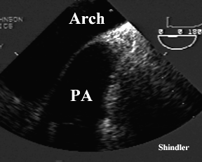
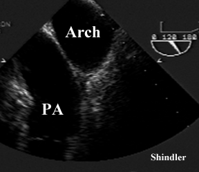
The aortic arch is usually scanned during transesophageal echocardiography while pulling back the probe and imaging the descending thoracic aorta. It is possible to measure the diameter of the arch, diagnose dissection and identify atherosclerotic changes. The normal direction of the aortic arch is also confirmed using the transverse imaging plane (zero degrees on the omniplane probe - as shown below). In most patients the interposed trachea limits imaging with the probe at this level. The two images below were obtained in a patient with an unusually well, simultaneously visualized, pulmonary artery. Changing the scanning plane to 120 degrees in the second image provided high definition images of the main pulmonary artery.
Back to E-chocardiography Home Page.
e-mail:shindler@umdnj.edu
The contents and links on this page were last verified on October 24, 2012
by Dr. Olga Shindler.