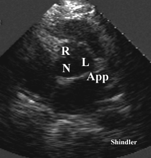
The left atrial appendage can be seen on some transthoracic adult studies.

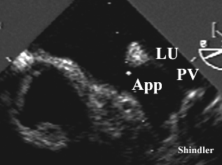
The left upper pulmonary vein is adjacent to the left atrial appendage. They are separated by a fold of tissue which can be quite visible on both transthoracic and transesophageal echocardiography. A persistent left superior vena cava occupies this space when it is present.
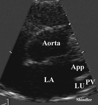
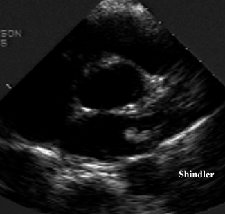
The tissue fold may resemble a mass on some transthoracic images as shown above, and indeed, quite often does so on transesophageal images, as shown below.
Original description by John Marshall:
On the development of the great anterior veins in man and
mammalia: including an account of certain
remnants of foetal structure found in the adult, a comparative
view of these great veins in the different mammalia, and an
analysis of their occasional peculiarities in the human subject.
Phil Trans R Soc Lond 1850;140:133-69
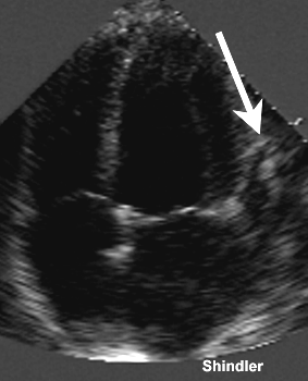 Unusually serpiginous left atrial appendage.
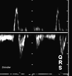 Normal Doppler flow pattern in the left atrial appendage.
There is appendage emptying following the P wave and
appendage filling following the QRS.
Back to E-chocardiography Home Page.

The contents and links on this page were last verified on November 18, 2012
by Dr. Olga Shindler