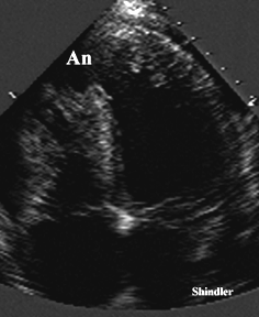
Cardiac involvement can be diagnosed with echocardiography. Left ventricular aneurysm, false aneurysm due to ruptured coronary artery, endomyocardial fibrosis, and diastolic filling abnormalities have been reported.

Large apical left ventricular aneurysm in a young patient with Behcet's.
J Vasc Surg 2001 Dec;34(6):1127-9
Behcet's disease: endovascular management of a ruptured peripheral arterial aneurysm. Kasirajan K, Marek JM, Langsfeld M.
Division of Vascular Surgery, University of New Mexico, School of Medicine, Albuquerque, NM 87131-5341, USA. kkasirajan@salud.unm.edu
Traditionally, bypass grafts are at a high risk for thrombosis or anastomotic degeneration in patients with Behcet's disease. We report the successful deployment of a vein-covered stent across the neck of a ruptured peripheral arterial aneurysm, via a remote site access, with intermediate-term follow-up. Covered stents may represent an attractive alternative to open surgical bypass for the management of aneurysms in patients with Behcet's disease.
Chest 2000 Aug;118(2):479-87
Intracardiac thrombus in Behcet's disease: a systematic review.
Mogulkoc N, Burgess MI, Bishop PW.
Department of Pulmonary Medicine, Ege University, Izmir, Turkey. 100046.1102@compuserve.com
BACKGROUND: Intracardiac thrombus formation is a rare but serious complication of Behcet's disease. We aimed to review the clinical and pathologic correlates of cardiac thrombus formation in the context of Behcet's disease. METHODS AND RESULTS: A comprehensive search of the medical literature was conducted using MEDLINE including bibliographies of all selected articles. Although the disease has a unique geographic distribution, being most common in the population of the ancient Silk Route, cases complicated by intracardiac thrombus have mostly originated from the Mediterranean basin and the Middle East. Young men appear to be most at risk, with the right heart the most frequent site of involvement. The first symptoms and signs of the disease frequently precede systemic organ manifestations. In those cases in which intracardiac thrombus occurs, it is apparent in more than half of cases on first recognition of the disease. CONCLUSION: A diagnosis of Behcet's disease should be considered if a patient presents with a mass in the right-sided cardiac chambers, even in the absence of the characteristic clinical features of the condition. This is particularly applicable if the patient is a young man from the Mediterranean basin or the Middle East.
Arch Mal Coeur Vaiss 2001 Apr;94(4):282-6
Endomyocardial fibrosis in Behcet's disease: a case report of a pseudo-tumoral form
Belmadani K, Dahreddine A, Benyass A, Hda A, Boukili MA, Ohayon V, Archane MI, Pavie A, Gandjbakhch I.
Service de medecine B, hopital militaire d'instruction Mohammed-V, Rabat, Maroc.
Endomyocardial fibrosis is very rare in Behcet's disease. The authors report the case of a 28 year old patient with Behcet's disease complicated by a pseudo-tumoral right ventricular formation on echocardiography. This misleading appearance suggested the diagnosis of cardiac thrombus or tumour and led to a surgical approach which revealed a fibrous moderator band suggesting endomyocardial fibrosis, confirmed by antomopathological analysis. Besides the originality of this case and the unusual pseudo-tumora l presentation, the authors underline the difficulties of establishing the diagnosis, despite the advances of medical imaging. The pseudo-tumoral intra-cardiac lesion in a suggestive clinical context (Behcet's disease) should raise suspicion of the diagno sis of endomyocardial fibrosis.
Heart 2001 Apr;85(4):E7
Behcet's disease with a large intracardiac thrombus: a case report.
Baykan M, Celik S, Erdol C, Baykan EC, Durmus I, Bahadir S, Erdol H, Orem C, Cakirbay H.
Department of Cardiology, Karadeniz Technical University Faculty of Medicine, 61080 Trabzon, Turkey.
Behcet's disease is recognised as a chronic multisystem disorder with vasculitis as its underlying pathological process. Cardiac involvement is rare and often associated with poor prognosis. A case of a 33 year old man with Behcet's disease, presenting with a large right ventricle and right atrial thrombus, is reported. Two dimensional (cross sectional), colour Doppler, and transoesophageal echocardiography, angiography, computed tomography, and magnetic resonance imaging were used to diagnose the disease. With cyclophosphamide and dexamethasone treatment, the cardiac lesions progressively resolved
Int J Card Imaging 2000 Oct;16(5):377-82
Cardiac thrombosis in a patient with Behcet's disease: two years follow-up.
Basaran Y, Degertekin M, Direskeneli H, Yakut C.
Kosuyolu Heart and Research Hospital, Istanbul, Turkey. mbasaran@sim.net.tr
A 28-year-old man with Behcet's disease was presented with cardiac symptoms in addition to previous complaints of oral and genital ulcers. A diagnosis of thrombosis was made and patient began to receive anticoagulant and immunosuppressive therapy and was followed by echocardiographic examination. Despite medical therapy, thrombosis recurred. Surgical excision was performed and histological findings were consistent with organizing thrombus. Nature of cardiac involvement and review of literature on cardiac thrombosis in Behcet's disease was discussed.
Intern Med 2001 Jan;40(1):68-72
Cardiac and great vessel thrombosis in Behcet's disease.
Ozatli D, Kav T, Haznedaroglu IC, Buyukasik Y, Kosar A, Ozcebe O, Dundar SV.
Hacettepe University Medical School, Department of Haematology, Ankara, Turkey.
Behcet's disease (BD) is a chronic relapsing systemic vasculitis in which orogenital ulceration is a prominent feature. The disease affects many systems and causes hypercoagulability. We present a 27-year-old male patient who exhibited widespread great vessel thrombosis including right atrial and ventricular thrombi in the setting of right-sided infectious endocarditis and orogenital aphthous ulcerations and erythema nodosum due to BD. We reviewed the enigmatic prothrombotic state of BD, and discuss our prior experiences in this field.
Back to E-chocardiography Home Page.

The contents and links on this page were last verified on November 5, 1998.