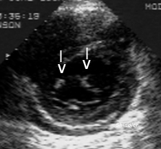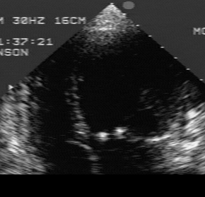

Adult with Down's syndrome and an av canal defect. In addition to a primum atrial septal defect and perimembranous ventricular septal defect, there is a cleft in the mitral valve. These two images portray the two dimensional echocardiographic appearance in the short axis parasternal and in the apical four chamber views. The mitral commisures appear symmetrically calcified. Doppler examination showed insufficiency without stenosis.

Back to E-chocardiography Home Page.

The contents and links on this page were last verified on January 13, 2000.