Semin Nephrol. 1995 Mar;15(2):87-105
Coarctation of the aorta.
Rao PS.
Department of Pediatrics, University of Wisconsin Medical School,
Madison, USA.
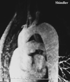
Coarctation of the aorta is an important and treatable cause of secondary hypertension. The prevalence of aortic coarctation varies from 5% to 8% of all congenital heart defects. Neonates and infants, especially when they have other associated cardiac defects, may present with signs and symptoms of heart failure. Children beyond infancy are usually asymptomatic and are most often diagnosed because of a murmur or hypertension on a routine examination. Palpation of the brachial and femoral pulses simultaneously will show decreased and delayed or absent femoral pulses. On measurement of blood pressure from arms and legs, a pressure difference of more than 20 mm Hg in favor of the arms may be considered as evidence for coarctation of the aorta. The coarctation can be demonstrated on suprasternal notch two-dimensional echocardiographic views along with increased Doppler flow velocity across the coarctation site. Cardiac catheterization shows significant peak-to-peak systolic pressure gradient across the coarcted segment, and aortography demonstrates the degree and nature of the aortic narrowing. Aortic coarctation may be relieved by surgery or by balloon angioplasty; in asymptomatic patients, therapy during the ages of 2 and 5 years is suggested. Surgical relief of coarctation may be achieved by resection and end-to-end anastomosis or by subclavian flap or prosthetic path angioplasty. Although results of surgery are generally good, there are some problems with the procedure, namely, mortality, morbidity and recoarctation, particularly in neonates and young infants and development of aneurysm, paraplegia, and paradoxical hypertension. Balloon angioplasty has been used by some cardiologists with resultant relief of obstruction, but concern for development of aneurysms and arterial complications remain. Although the immediate results for surgical or balloon therapy for isolated coarctation are good, long-term prognosis is largely undetermined. Limited long-term follow-up studies suggest significantly lower survival rates compared with normal population; age at intervention and the degree and duration of hypertension before intervention may affect long-term survival.
Pediatrics. 1996 Sep;98(3 Pt 1):378-82
Early diagnosis of coarctation of the aorta in children:
a continuing dilemma.
Ing FF, Starc TJ, Griffiths SP, Gersony WM.
Department of Pediatrics, Columbia University, College of
Physicians and Surgeons, New York, New York, USA.
Because late repair of coarctation of the aorta (COA) is associated with premature cardiovascular disease in adult life, early detection and treatment is important. OBJECTIVES: To determine the timing of referral to see whether early detection of COA has improved in the past decade, to evaluate the pattern of and reasons for medical center referral, and to assess the clinical signs relating to the diagnosis of COA. METHODS: The records of 50 consecutive patients older than 1 year who had surgical repair of COA from 1980 to 1990 were reviewed. The age of referral, pattern of referral, and presence of standard clinical signs of COA were analyzed, and data were compared with those from the previous decade. RESULTS: The mean and median ages at referral were 8.4 and 5.8 years, respectively. Pediatricians accounted for 64% of the referrals. A specific diagnosis of COA was made in 2 (4%) of 50 patients before referral to a pediatric cardiologist. The most consistent clinical findings were a cardiac murmur and a systolic blood pressure gradient between the arms and legs of greater than 10 mm Hg, which were both present in all patients. Lower-extremity pulses were decreased in 37 (74%) and absent in 9 (18%). Forty-seven children (94%) had upper-extremity hypertension (> 95th percentile for age); 25 (50%) had systolic blood pressure higher than 140 mm Hg. COA would have been missed in 82% of children if absent lower-extremity pulses were required as a diagnostic feature. These findings were similar to those reported by our institution in the previous decade, suggesting that early detection has not improved. CONCLUSIONS: The timing of, reasons for, and sources of referral for COA in this study, compared with data from the previous decade, indicate no improvement in early detection of COA by pediatricians. Screening all children for COA by routinely measuring upper- and lower-extremity blood pressures during at least one physical examination after the newborn period is mandatory.
Ann Dermatol Venereol. 2000 Mar;127(3):292-5
Facial hemangioma associated with arterial anomalies, coarctation
of the aorta, and eye abnormalities: PHACES syndrome
[Article in French]
Buzenet C, Hamel-Teillac D, Acar P, Becquet F, Curan D,
Michaud V, Sidi D, De Prost Y.
Service de Dermatologie, Hopital Necker-Enfants Malades,
149 rue de Sevres, 75015 Cedex.
BACKGROUND: Hemangiomas are frequent in childhood. Their association with dysmorphic anomalies is rare. Recently, the acronym "PHACES syndrome" was proposed to emphasize the association of Posterior fossa malformations, Hemangiomas, Arterial anomalies, Coarctation of the aorta and cardiac defects, Eye abnormalities, and Sternal malformations. CASE REPORT: A female child, 3 months old, had a large facial hemangioma. The physical examination was normal otherwise. A choroidal hemangioma and a papillary abnormality, causing amblyopia, were detected. The brain magnetic resonance imaging was normal. A subglottic hemangioma was found at endoscopy. At the age of 16 months, physical examination disclosed a heart murmur and coarctation of the aorta was detected. Moreover, the cardiac angiography showed diffuse arterial lesions. Strict surveillance was decided as there were no manifestations. DISCUSSION: Different abnormalities have been described to be associated with large facial hemangiomas. Frieden has grouped these abnormalities under the acronym PHACES. She described 43 hemangiomas and found 74 p. 100 Dandy Walker malformations and other posterior fossa malformations, 41 p. 100 arterial anomalies, 26 p. 100 cardiac or aortic malformations, 23 p. 100 ophthalmologic abnormalities. There is a high risk for the hemangiomas to develop in an airway localization. The prevalence of facial hemangiomas associated with other malformations is, to our knowledge, not known. In our department, 56 children were treated with corticosteroid therapy for severe facial hemangioma. 11 p. 100 had a cerebral abnormality. There were no cases with cardiac malformation or dysmorphism. PHACES syndrome is very rare but easy to remember. Thus in patients presenting a large facial hemangioma, it is important to conduct an attentive neurological examination completed by brain imaging and an extensive cardiovascular exploration. Special attention should be given to the ophthalmologic and sternal examinations as well as the search for hemangiomas in an airway localization.
Back to E-chocardiography Home Page.
e-mail:shindler@umdnj.edu
The contents and links on this page were last verified on December 31, 2003.