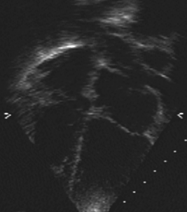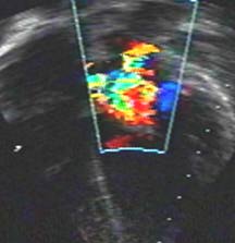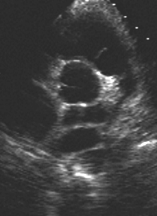
Surgically corrected total anomalous pulmonary venous drainage. The four pulmonary veins converge behind the left atrium into a common pulmonary vein. The surgery consisted of creating a communication between the common vein and the left atrium. This is shown by the color flow image with laminar (yellow) flow in the common vein. There is a narrowing of the flow with some turbulence at the junction to the left atrial cavity (where the color spreads out).


Short axis view in the same patient showing the common vein behind the left atrium.

Echocardiographic differentiation between cor triatriatum and supravalvular mitral ring:
Cor triatriatum membrane is more curved, may have windsock contour, moves toward the mitral valve plane in diastole, all pulmonary veins drain proximal to the membrane, left atrial appendage and foramen ovale are distal to the membrane.
Supramitral ring attaches to the base of the mitral valve past the left atrial appendage and past the foramen ovale, it moves away from the mitral valve in diastole, mitral leaflet motion is abnormal with prolonged Doppler pressure half time.
Back to E-chocardiography Home Page.
e-mail:shindler@umdnj.edu
The contents and links on this page were last verified on September 20, 2002.