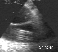
Echocardiographic appearance of a Denver shunt in the superior vena cava.
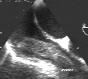
Right atrial mass originating at a Denver shunt in the superior vena cava. Histology showed a fibrin thrombus with numerous aggregates of of embedded neutrophils.
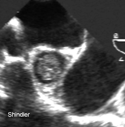
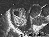
Cross section of the thrombus in the superior vena cava.
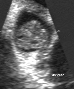
Closeup of the same thrombus.
Denver shunt thrombus in the right atrium.
Gosse P, Guez S, Roudaut R, Deville C, Dallocchio M. reported
a similar complication of the Le Veen shunt: a right atrial
ventricular chamber pseudotumor.
(Clin Cardiol 1987 May;10(5):370-1)
A right atrial-ventricular chamber thrombus was
discovered four years after insertion of a Le Veen shunt for
treatment of refractory ascites. A two-dimensional
echocardiogram, performed after the discovery of an isolated
systolic murmur, demonstrated a tumorlike mass seated in both
the right atrium and the right ventricle. The mass was surgically
removed and histologic examination confirmed that it was a
thrombus that developed at the tip of the catheter. The authors propose
that two-dimensional echocardiography should be performed periodically
for the survey of such intracardiac devices.
Back to E-chocardiography Home Page.

The contents and links on this page were last verified on September 6, 2005.