 Right atrial thrombus (4).
Dr. Victor Grech
Paediatric Senior Registrar
Paediatric Department
St. Lukes Hospital
Malta
Ms. Rachel Danvers
Chief Echocardiography Technician
Cardiothoracic Unit
Great Ormond Street Hospital for Children NHS Trust
London WC1N 3JH
UK
Corresponding Author
Dr. Victor Grech
Surface mail address - as above
e-mail:
victor.e.grech@magnet.mt
Introduction
Echocardiography has revolutionised the practice of paediatric
cardiology by allowing non-invasive diagnosis and follow-up of a
wide variety of conditions, particularly congenital heart disease
(CHD) - allowing patients to be diagnosed, followed-up and even operated
without invasive investigations (1,2). Echocardiography machines have
become highly sophisticated and versatile pieces of equipment. This
article illustrates non-CHD applications of echocardiography in the
setting of a tertiary paediatric cardiac referral centre.
Methods
Images were obtained by Acuson XP-10, Acuson Sequoia and Toshiba SSH654
machines from patients referred to the Cardiothoracic Unit, Great Ormond
Street Hospital for Children NHS Trust. All graphics were downloaded to
conventional photographic paper, and then converted to digital images using a
Hewlett Packard Scanjet IICX. The images were annotated using Paint Shop Pro.
Cropping and contrast adjustments were made using Adobe Photoshop.
Intracardiac masses
Right atrial thrombus (4).
 Wilm's tumour with extension to right atrium (5).
Extracardiac intracirculatory masses
 Thrombus formation in the innominate vein after bidirectional
Glenn procedure for tricuspid atresia (6).
Thrombus formation in the innominate vein after bidirectional
Glenn procedure for tricuspid atresia (6).
Effusions
 Pericardial effusion with fibrin formation (7).
 Bilateral pleural effusions (8).
Localisation of central lines and associated thrombi/vegetations
 Umbilical arterial line in descending aorta seen both in short axis
and long axis aortic views (9).
Umbilical arterial line in descending aorta seen both in short axis
and long axis aortic views (9).
Ventriculo-arterial shunt
 Infected ventriculo-arterial shunt with vegetation formation at
tip of shunt in right atrium (10).
Infected ventriculo-arterial shunt with vegetation formation at
tip of shunt in right atrium (10).
Extra-corporeal life support cannulae
 Extra-corporeal life support venous cannula in
superior vena cava extending to the right atrium.
Extra-corporeal life support venous cannula in
superior vena cava extending to the right atrium.
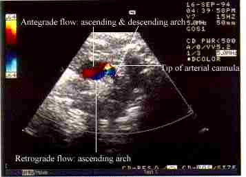 Arterial cannula in apex of aortic arch (11).
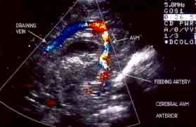 Arteriovenous malformations causing heart failure
due to extracardiac left-to-right shunting (12)
Arteriovenous malformations causing heart failure
due to extracardiac left-to-right shunting (12)
Diagnosis of post-operative diaphragmatic paralysis.
Unilateral and even bilateral, partial or complete diaphragmatic paralysis due to trauma to the phrenic nerve, is an occasional complication of surgery for CHD. Upward and downward movement of the diaphragm in expiration and inspiration respectively, can be screened in real-time both ultrasonographically and fluoroscopically. The left and right components of the diaphragm can be viewed simultaneously from the subcostal window by US allowing paradoxical movement to be visualised in ventilated patients with unilateral diaphragmatic paralysis (13).
Discussion
Cardiac applications of ultrasonography techniques have undergone
tremendous strides since the inception of M-mode
echocardiography. 2-dimensional echocardiography allows detailed
intracardiac analysis of complex malformations, and the
availability of pulse-wave Doppler and continuous-wave Doppler
since the mid-1980s and colour Doppler since the late 1980s has
allowed even more information to be gathered in the cardiac
ultrasound examination (14). Full diagnosis, follow-up and even
surgery of patients with CHD can be undertaken without resort to
more invasive techniques in many individuals (1,2).
The images in this article illustrate how chocardiography has been utilized for the diagnosis and/or monitoring of non-congenital heart problems in a tertiary pediatric referral hospital (15).
Abstract on the use of echocardiography in a Canadian level three neonatal intensive care unit.
Back to E-chocardiography Home Page.
e-mail:shindler@umdnj.edu