

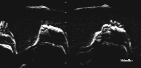
Large mobile calcified vegetation attached on the left atrial side of the mitral annulus. M mode below shows the variable appearance of the vegetation surface from beat to beat. The patient has chronic renal failure.
Transthoracic and transesophageal images of a vegetation on the wire of an implanted cardioverter defibrillator.
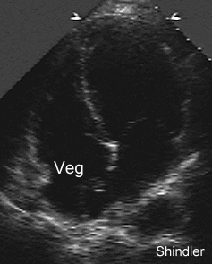
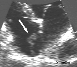
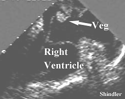
Destructive abscess with calcification in a young patient with end stage renal failure.
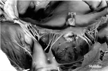 Fibrocalcific nodular protrusions (FCN) on both sides of the mitral
valve projecting into the left atrium as well as into
the left ventricle.
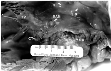 Right atrial fibrocalcific nodule (RAN) located just
above the tricuspid valve (TV). Ruptured, thickened
chordae tendineae (CT). Septal abscess (SA).
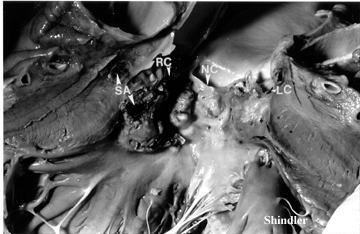 Calcification of the aortic leaflets: RC, NC and LC are
the right, non, and left coronary cusps respectively.
Abscess of the interventricular septum (SA).
Back to E-chocardiography Home Page.
e-mail:shindler@umdnj.edu
The contents and links on this page were last verified on May 30, 2002.