Gallavardin Phenomenon and Echocardiography
Case Presentation
A 75 year old male presents with progressive dyspnea on
exertion. He denies any chest pain. One year ago he had
a syncopal episode, but did not seek medical attention.
He does not see a physician regularly. He is not sure
whether he was told that he has a murmur in the past.
On auscultation there is a harsh ejection type systolic
murmur in the second right intercostal space. It is also
audible in the right carotid region. There is a musical
murmur at the cardiac apex. It does not radiate to the
left axilla. Both murmurs decrease with handgrip. You
request an echocardiogram with Doppler looking for the
diagnosis of combined aortic stenosis and mitral regurgitation.
The echo report shows severe aortic stenosis, but to your
surprise there is no mention of mitral regurgitation.
Discussion
The diagnosis of aortic stenosis (AS) is made with auscultation.
The systolic murmur of AS is usually best heard at the upper
right sternal border and it typically radiates to the neck.
It is an ejection type murmur with a crescendo-decrescendo pattern. The
duration of the murmur is variable as is the peak intensity. A late
peaking murmur is consistent with severe AS. An early peaking murmur
is consistent with mild AS.
One auscultatory task is to distinguish the murmur of AS from that
of mitral regurgitation (MR). The murmur of MR is usually best
heard at the apex, but Gallavardin described a dissociation of the AS
murmur into two components. The first component is the typical harsh
systolic right sternal border murmur that radiates to the neck. It
is due to the high velocity AS jet in the ascending thoracic aorta.
The second component mimics MR. It is best heard at the apex. This
apical component of the aortic stenosis murmur is musical. It
therefore, cannot be easily distinguished from MR. It is presumably
due to high frequency vibrations traveling to the apex from the
calcific aortic valve. Maneuvers during auscultation are useful.
Handgrip increases resistance to arterial forward flow and facilitates
regurgitant reverse flow. It therefore, increases the murmur of MR and
decreases the intensity of the AS murmur. An unusually long cardiac
cycle will cause an AS murmur to increase in intensity, whereas the
MR murmur will remain unchanged. This can be heard during a compensatory
pause following a premature ventricular contraction, or during a long
cardiac cycle in atrial fibrillation.
AS is quantitated and followed using transthoracic echocardiography.
Thin, normally moving aortic leaflets exclude the presence of valvular
AS. Thick aortic leaflets can be stenotic, or just sclerotic. A peak
gradient greater than 25 mm Hg is used in our lab as the criterion
for presence of stenosis rather than sclerosis. The peak and mean
gradients across the valve can be determined non-invasively with
Doppler. The area of the stenotic aortic valve orifice is calculated
by multiplying the area of the left ventriclular outflow by the ratio
of the left ventricular outflow gradient to the stenosis gradient.
Both gradients are easily measured with Doppler and can be used for
non-invasive follow-up of the progression of AS. Echocardiography also
shows the degree and distribution of left ventricular hypertrophy.
However, when it comes to distinguishing whether the etiology of the
murmur is AS or MR, it is color Doppler that clarifies the nature of
the systolic murmur by showing the presence or absence of mitral
insufficiency. Therefore, in the absence of color Doppler evidence
of MR, the systolic murmur can be ascribed to the thick, or calcified
aortic valve.
Unfortunately, in a large number of patients MR coexists with AS.
There are many reasons for this. Progressive left ventricular hypertrophy
in patients with progressive aortic stenosis is considered an adaptive
response to prevent ventricular dilatation and progression of MR.
When MR does progress along with the AS, it is a maladaptive consequence.
Patients with aortic stenosis may have associated coronary artery
disease. MR can occur following inferior myocardial infarction because
of disrupted leaflet coaptation. Chordal rupture (rather than the
catastrophic papillary muscle rupture) can cause different grades of
MR. Left ventricular dilatation following an anterior myocardial
infarction can involve the mitral annulus and cause MR. Patients
with AS have variable degrees of left ventricular hypertrophy.
Typically the hypertrophy is concentric with variable cavity to
wall thickness ratios. Some patients may have an intracavitary
gradient. Left ventricular ejection may result in intraventricular
obstruction, which in turn, can cause MR. In the patient with
calcific aortic stenosis, there is often calcification of the mitral
annulus as well. This results in some disruption of mitral leaflet
coaptation with consequent mitral insufficiency.
Although color Doppler can be very helpful in excluding the presence
of MR, it can be misleading about the severity of MR when it coexists
in patients with AS. Color Doppler is simply a spatial display of
velocities. MR Doppler velocities are always high because there is
always a large gradient between the left ventricle and left atrium
in systole. Yet, when they are displayed two dimensionally with
color Doppler, MR color flow velocities can both overrepresent and
underrepresent the actual severity of the MR. In patients with
severe AS there is an extremely high left ventricular systolic
pressure which can make hemodynamically moderate MR appear severe
on color Doppler.
Assessment of the mitral valve is important in the patient undergoing
valve replacement for AS. The assessment of MR consists of two-dimensional
and M-mode visualization of the mitral valve followed by evaluation
of the color Doppler flow pattern. It then proceeds to Doppler
evaluation of the pulmonary veins. Typically, the patient undergoing
aortic valve replacement for AS will not require mitral valve replacement.
The decision to repair or replace the mitral valve in an AS patient
has to be made before starting the operation, since the surgeon prefers
to operate on the mitral valve before operating on the aortic valve.
Preoperative transesophageal echocardiography may be necessary to
resolve this question because it can display pulmonary venous inflow
with greater detail. It also provides better images of the mitral
leaflets and chordae. After the stenotic aortic valve is replaced,
the degree of MR on color Doppler is decreased because the left
ventricular systolic pressure is decreased.
A systolic murmur is a valuable clinical finding that needs to be
investigated with careful auscultation that includes hemodynamic
maneuvers. Echocardiography complements auscultation by resolving
some of the pitfalls.
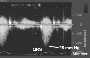 Continuous wave Doppler measurement of aortic valve
gradient. The peak gradient is calculated as four
times the square of the peak velocity. Ealy peaking
flow in this patient has the same implication as an
early peaking murmur - mild aortic stenosis.
Continuous wave Doppler measurement of aortic valve
gradient. The peak gradient is calculated as four
times the square of the peak velocity. Ealy peaking
flow in this patient has the same implication as an
early peaking murmur - mild aortic stenosis.
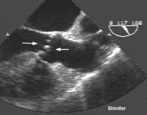 Transesophageal echo view of a calcified aortic
valve (arrows).
Transesophageal echo view of a calcified aortic
valve (arrows).
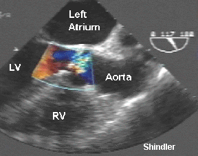 Color flow of the stenotic aortic valve.
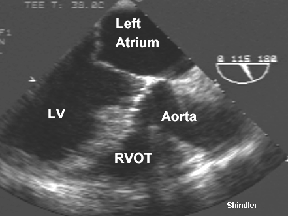 Transesophageal echo view of the mitral valve.
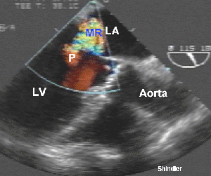 Color Doppler of mitral regurgitation during the
isovolumic filling period (aortic valve is still
closed). MR denotes the regurgitant jet in the left
atrium. P is the proximal isovelocity flow convergence
used for quantitation of the degree of MR.
Color Doppler of mitral regurgitation during the
isovolumic filling period (aortic valve is still
closed). MR denotes the regurgitant jet in the left
atrium. P is the proximal isovelocity flow convergence
used for quantitation of the degree of MR.
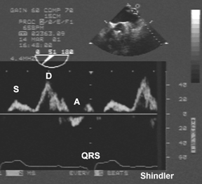 Left upper pulmonary vein pulsed wave Doppler flow
pattern obtained by transesophageal echocardiography.
There are three waves: systolic S wave, diastolic D
wave, atrial reversal A wave. This image shows an
abnormal diastolic dominant pattern. Normally the S
wave is larger than the D wave. In severe MR there
may be another wave of systolic flow reversal.
Left upper pulmonary vein pulsed wave Doppler flow
pattern obtained by transesophageal echocardiography.
There are three waves: systolic S wave, diastolic D
wave, atrial reversal A wave. This image shows an
abnormal diastolic dominant pattern. Normally the S
wave is larger than the D wave. In severe MR there
may be another wave of systolic flow reversal.
Back to E-chocardiography Home Page.

The contents and links on this page were last verified on
October 31, 2006.