THE SIGNIFICANCE OF A HITHERTO UNDESCRIBED WAVE IN THE JUGULAR PULSE.
By ALEXANDER G. GIBSON, M.A., MB. Oxon., M.R.C.P. Lond.
The Lancet Nov 16, 1907
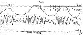
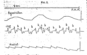
In the course of a study of jugular pulses in normal persons I have noticed that in those whose pulse-rate is slower than the average (60 per minute or under) a wave between the "v" wave and the "a" wave is often to be seen. It is most typically seen in normal young adults who are in the habit of taking vigorous exercise or who are convalescent from acute diseases. Such waves are seen in Figs. 1 and 2 and marked "b". The form of the wave varies; sometimes it is an almost vertical uprising with a straight top, at other times it is a well-defined short wave. The form of wave seen in its largest examples is one in which on the upstroke there is a small indentation just before the summit, and is similar in many respects to another nick which has been described on the "v" wave as being due to the closure of the semilunar valves. When a series of these waves are shown in a tracing they are found to bear a constant relation to the preceding systole as measured from the beginning of the auricular or carotid wave. This is well seen in Fig. 1 where owing to a marked respiratory variation in the heart rate the interval between the "b" and succeding a wave is seen to increase with the slowing of the pulse. The following table shows the time interval elapsing between the "c" and this wave in some cases of which good records have been obtained:-
Condition: Normal; "c" "b" interval: 0.56 - 0.66 second
Condition: Normal; "c" "b" interval: 0.64 second
Condition: Convalescent from influenza; "c" "b" interval: 0.5 -0.6 second
Condition: Convalescent from pneumonia; "c" "b" interval: 0.6 - 0.64 second
Condition: Convalescent from rheumatic fever; "c" "b" interval: 0.52 - 0.6 second
The variation in the "c" "b" interval is probably partly mechanical from the differences in the fullness of the jugular vein. The "c" "b" interval sometimes bears a relation to the respiration but the differences are so slight that they would easily fall within the limits of error, and the variation is not always in the same direction in two individuals.
In the jugular pulse thanks to the researches of Mackenzie and others we have in the small nick on the upstroke of the "v" wave a point where systole ends, this being the position of the closure of the semilunar valves. The period, therefore, which intervenes between this and the subsequent auricular wave both the cavities of the heart are in diastole. During this period the "b" wave occurs. In pulses of average rate, between 70 and 80, the interval is so short that any wave that may be present is not distinctly visible, but in hearts of slower rate, as can be seen from the tracings, it is in some instances extremely well marked. The events which occur in the heart during this period are: first, the replacement of the heart after its protrusion during systole, probably causing the main part of the "v" wave; and secondly, the emptying of the veins and auricle into the ventricle which becomes gradually filled. Without examining the evidence in favour of the view it is accepted, I believe, that when the ventricle becomes moderately full the mitral and tricuspid valves float up by reason of eddies behind them in the ventricle, and the closure of the aperture by this means prevents regurgitation. An increase of pressure in the ventricles now causes these free edges to be closely pressed together in apposition. Apart from the filling of the ventricles, auricles, and veins which goes on during diastole and may cause an overflow wave in the jugular vein, this closure, at least in slowly-beating hearts with ventricles of normal size, must occur some time before the auricle begins to contract, for, if not, one must conceive either a slow filling of the ventricle for a slowly beating heart or an enlarged ventricle. It is conceivable then that this extra wave may be due to the closure of the auriculo-ventricular valves by the natural filling of the ventricle with blood and the slight increase in pressure which the upward movement of the valves would cause in the venous system. Here it may be pointed out that the form of the wave suggests a mechanism of this kind, for no mere overflow wave such as could be caused by a gradual filling up of the ventricles, auricles, and veins alone could cause the nick which has been noticed as characteristic of many examples of the wave.
In two of the cases recorded in which the heart sounds were carefully studied it was possible to detect a third sound between the second sound and the succeeding first sound. It was heard only at the apex in one, while in the other case it was heard at the apex and over the jugular pulsation in the neck. The sound is by no means easy to hear; extraneous sounds must be eliminated as far as possible and special attention paid to the interval in which the sound is expected. Nor is it possible to hear the sound at every systole; from my own observation it is best heard in the short interval between expiration and inspiration. The quality of the sound is low-pitched and clear, with no suspicion of harshness or anything suggesting a murmur. Except for the pitch it recalls in type the second sound as heard in fat persons. It is slightly increased in intensity and raised in pitch by pressure on the abdomen. It was desirable to see if any relation existed between the time of the sound and that of the wave; accordingly, failing apparatus for recording the sounds of the heart graphically, an attempt was made to record the time when the sound was heard, or rather expected, on the same tracing as the jugular pulsation was recorded. This method has not been sufficiently successful to record the results except in one instance which will be referred to. Professor Einthoven who has recently been engaged in the recording of heart sounds by means of his string galvanometer (Saiten-galvanometer) has been good enough at my request to look over his tracings of normal subjects and his letter convinces me that the sound and wave are due to the same cause. He writes to say that in one case investigated an extra sound was noted at the apex after the second sound. The subject had a pulse of 67. In two tracings of the sounds in this case which Professor Einthoven sends me the interval between the beginning of the first sound and that of the extra sound varies between 0.44 - 0.48 sec. As in the observations by means of the stethoscope, the sound is occasionally not recorded. If it is considered that Professor Einthoven's instrument probably records some of the auricular sound previous to the first sound and that from the table given previously the time interval from carotid wave to "b" wave varies somewhat in different individuals, the agreement between Professor Einthoven's and my own observations is striking. Moreover, I have been in the habit of taking as the beginning of the "b" wave the place where the nick is found on the upstroke, and this again would lessen the "c" "b" interval to 0.5 of a second.
It is highly probable, therefore, that the sound and the wave in the regular pulse are due to the same cause. There are three possibilities for the explanation of a sound in the diastolic period; first, a muscular sound having to do with something other than auricle or ventricle; secondly, a rush of blood through an orifice; and thirdly, the tension of a membrane. Except the venous musculature no muscular contraction in this period is conceivable, and this is of such small proportions that it would not be likely to produce an audible sound; moreover, the "b" wave bears no relation to the succeding auricular systole which we should expect to find were this the cause. The second possibility is again unlikely, for everything that is known of the filling of the heart, at least in normal persons points to a very gradual process. This is not so, however, with the third hypothesis. By observation on the excised heart it can be seen that as the ventricular cavity fills the auriculo-ventricular valves float up and close automatically when the ventricle is full, and it is easy to imagine that the inrush of blood would be followed by a reaction on the ventricular walls in proportion to the momentum of the inrush, so that with a rapid and full stream the tightening of the valves would be quick and strong while with a slow stream the process would be more gradual yet in no wise less complete. The fact that this explanation necessitates the closure of the valves before the auricular beat is no objection; the amount of force necessary to push open the valves is extremely small and the heart would be the more rather than the less efficient pump because of the double closure of the valves. The sequence of events in the diastolic period would then be as follows. After the replacement of the heart which follows systole and causes the "v" wave in the jugular pulse the blood flows into the ventricle, the valves float up, gradually when the venous pressure is low, quickly when it is high, and the orifice closes; the auricle is now filled by blood from the veins; it contracts and forces the greater part of its contents into the moderately distended ventricle which then becomes fully so, the valves close again from the increase of pressure set up in the ventricle by the auricular contraction; finally, the ventricular contraction keeps the valves closed till the end of systole.
Boyd inquiring into the cause of the reduplication of the second sound, heard at the apex only, in cases of mitral stenosis, allocates the formation of the sound to the mitral valve but considers it to be a presystolic murmur produced by flow of blood through a constricted orifice. In testing the capacity of the pulmonary valves to produce a sound on closure he found a head of six inches of water was necessary for the sound of the tension of the valves to be audible by the phonendoscope. Sansom discussing the causation of the same phenomenon, considers it due to the sudden release, by diastole, of the blood in the auricles and veins, and the forcible inflow into the ventricle followed by sudden concurrence and tension of the valve cusps. It is not possible as yet to decide whether the right and left side of the heart participate equally in the causation of both the wave and the sound; the presence of the wave in the jugular pulse would suggest its causation in the tricuspid valve, while the localisation of the sound at the apex heat would suggest as its site the mitral. This, however, is not of great importance to the main proposition.
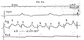
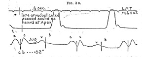
It is to be supposed that if the explanation of these observations is correct, the wave would alter in its time relations with a variation in the size of the cavities of the heart. This is found to be borne out by observation. It is admitted that in pneumonia there is some affection of the heart muscle even in previously healthy people, probably in the direction of an impairment of contractibility and tonicity. Fig. 3 A is the record of the apex beat, jugular pulse, and radial the first day after the subsidence of fever in an otherwise healthy young adult after a sharp (four days) attack of pneumonia. The "c" wave is seen to he separated frem the "b" wave by an interval of 0.6 sec. In Fig. 3 B, taken four days later, is seen the time of the third (extra) sound in relation to the jugular pulse. It is seen that this sound, as marked, begins at the top of a wave which immediately succeeds V, probably. the "b" wave; but if so, this is now only separated from the preceding a wave by 0.52 sec. A second case of mitral and tricuspid failure with oedema, a slow pulse the result of digitalis, and apex beat two inches outside the nipple line showed in one tracing a "c" "b" interval of 1.06 sec., in a tracing taken four weeks later, when the apex beat was in the nipple line, gave a "c" "b" interval of 0.74 sec. At the time of the latter tracing it was not possible to hear any extra sound either at the apex or in the neck.
Since writing the above Dr. James Mackenzie has directed my attention to a paper by Hirschfelder in the Johns Hopkins Bulletin, June-July, 1907, in which tracings from a patient with myocardial disease are given showing a wave similar in form and relation to that described in this paper. Hirschfelder found the same wave in the normal individual, and in this but not in the former case he was able to hear over the tricuspid area at the end of expiration a third heart sound simulating the protodiastolic gallop rhythm. The floating-up of the mitral and tricuspid valves is given as a possible cause, but no conclusions are formed.
Oxford.
Back to E-chocardiography Home Page.
e-mail:shindler@umdnj.edu