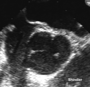
Lambl (Wein Med Wschr vi, 1856, 244) described small filiform processes on the aortic valve. Margerey (J Path Bact 1949; 61:203-208) studied 250 mitral valves and postulated that the mechanism of formation is intimal damage due to mechanical trauma at the leaflet coaptation.
The damaged area is covered by fibrin, which subsequently becomes uplifted, or partially detached from the valve surface. A layer of intimal cells covers the surface of the fibrin deposit. The enclosed fibrin becomes condensed and hyaline constituting the excrescence. Organized hyaline substance is later replaced by fibrous and elastic tissue.
Morphologic characteristics and structure of surface excrescences
(Lambl's excrescences) in the normal aortic valve.
Hurle JM; Garcia-Martinez V; Sanchez-Quintana D
Am J Cardiol 1986 Dec 1;58(13):1223-7
The incidence, morphologic characteristics and structure of surface valve excrescences (Lambl's excrescences) were studied by transmission and scanning electron microscopy of the aortic valve of 56 human subjects, age range birth to 91 years, without cardiac disease. Valve excrescences consisting of a core of connective tissue covered by the endocardium were observed in 90% of the subjects; the incidence was significantly lower in patients in the first decade of life. Two types of excrescences, lamellar and filiform, were found. Lamellar excrescences are located along the lower boundary of the lunulas and occurred more often in those younger than 30 years. Filiform excrescences appear most often in the nodulus Arantius and in the free- margin of the cusps. The excrescences of the nodulus are the most numerous. Free-margin excrescences are the least numerous and occur more frequently in persons older than 40 years. The connective tissue core of the filiform excrescences contains abundant collagen fibrils and elastic material arranged in apposed layers with different collagen fibril orientation. A circular zone devoid of identifiable connective tissue is present at the center of the filiform excrescences.
Back to E-chocardiography Home Page.

The contents and links on this page were last verified on November 18, 2012
by Dr. Olga Shindler