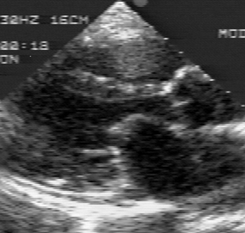
The parasternal, long axis view is the initial echocardiographic imaging approach in most adult patients. When the diagnosis is congestive heart failure, it provides immediately useful information and guides the subsequent course of echocardiographic image acquisition.
Once the parasternal long axis image is optimized, one may begin by scrutinizing the aortic valve. The presence of aortic valve calcification prompts a systematic evaluation for aortic stenosis severity. The presence of heart failure in a patient with aortic stenosis is an ominous finding often prompting urgent open-heart surgery to replace a severely stenotic aortic valve. Hence the echocardiographic abnormalities of aortic valve calcification, perhaps aortic valve doming, and certainly Doppler evidence of turbulent flow, will warrant complete evaluation of the gradient across the aortic valve. The presence of decreased systolic left ventricular function can alter the reliability of Doppler in assessing the true gradient across the aortic valve.
The next point of attention in the parasternal long axis view is usually the mitral valve apparatus. The classic mitral stenosis hockey stick appearance of the anterior mitral leaflet is rare, but can clarify the clinical diagnosis of congestive heart failure. The dyspnea is secondary to pulmonary congestion but systolic left ventricular function may well be normal.
The most common abnormality on color Doppler related to the patient with congestive heart failure may be some degree of mitral insufficiency. Mitral insufficiency can be either primary or secondary to the heart failure. Primary mitral insufficiency resulting in heart failure needs to be evaluated for etiology. This can become apparent in some patients in the parasternal long axis view immediately after placing the transducer on the chest. For example, the patient can have mitral insufficiency secondary to mitral valve prolapse .
Secondary mitral insufficiency in the patient with congestive heart failure may be due to a dilated left ventricle that has caused dilatation of the mitral annulus with alteration of the mitral leaflet coaptation and some degree of mitral insufficiency.
Conversely, a commonly overlooked cause of heart failure, especially in older patients, is not a systolic problem, but rather a diastolic problem. In diastole, filling may be impaired in elderly patients with noncompliant left ventricles. Often, there will be calcification of the mitral annulus and chordae. The combination of left ventricular hypertrophy, small left ventricular internal dimensions, mitral annulus calcification (resulting in systolic disruption of the mitral leaflet closure and some degree or mitral insufficiency) is quite commonly seen in the elderly patient with a stiff, noncompliant left ventricle where the congestive heart failure is due to diastolic rather than systolic dysfunction.
The segmental analysis of left ventricular wall motion begins
in the parasternal long axis view. Scrutinizing the proximal
portion of the intraventricular septum may reveal the
presence of hypokinesis, akinesis, dyskinesis or a scar.
Such patients may have a stenotic lesion in the very
proximal portion of the left anterior coronary artery or
possibly in the left main coronary artery. This is an
ominous finding.
Patients with circumflex coronary artery disease or right
coronary artery disease may exhibit motion abnormalities in
the posterior left ventricular wall. The basal and mid
portions of this wall are seen in the parasternal long axis
view.
A rare but important cause of symptoms of congestive heart failure is idiopathic hypertrophic subaortic stenosis. This is a rare entity, but can be autosomal dominant and therefore can be present in many members of a single family. In addition to the basal septal hypertrophy, abnormal ultrasound reflections (due to the associated myocardial fiber disarray) may be present. Other findings available on the parasternal long axis view are systolic anterior motion of the mitral leaflets and chordae as well as mid-systolic closure of the aortic valve.
The presence of pericardial fluid can also be established in the initial parasternal images. The size of the effusion can be estimated. Hemodynamic compromise can be demonstrated in this view by looking for diastolic flattening of the right ventricular free wall.
Needless to say, left ventricular systolic and diastolic chamber sizes, as well as wall thickness, are measured in this view to quantitate left ventricular function.
These examples illustrate the value of the initial parasternal long axis images for the echocardiographic examination of a patient with a the diagnosis of heart failure. The first impressions formulated here are well on their way to providing the correct diagnosis in this important clinical entity.
Return to E-chocardiography Home Page.
E-mail: shindler@umdnj.edu
The contents and links on this page were last verified on April 15, 2005.