Lipomatous Hypertrophy of the Atrial Septum
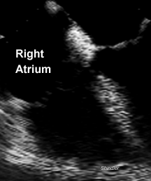
Fatty infiltration of the atrial septum with sparing of the
membrane of the fossa ovalis.
Echocardiographic Criteria for Diagnosis
- Bilobed appearance as in the above image.
- Septal thickness of 15 mm or more.
- Exclusion of other causes of atrial septal thickening, such as
amyloidosis, surgical patches, tumors.
References
Maillet-Vioud C, Eicher JC, Falcon S, Delescaut M, Gomez MC,
Cottin Y, Brunotte F, Brenot R, Louis P, Wolf JE. Lipomatous
septal hypertrophy. Apropos of 3 cases.
Ann Cardiol Angeiol (Paris) 1994 Jun;43(6):328-30.
Lipomatous hypertrophy of the inter-atrial septum is
characterised by fatty accumulation in the inter-atrial septum.
Long unrecognised, it has been discovered anew by virtue of the
use of transesophageal echocardiography. The authors report three
cases, including one presenting as acute ischemia of the right
lower limb. After studying the contribution of imaging techniques
to its diagnosis, the authors consider the principal differential
diagnoses of this condition.
Kozelj M, Angelski R, Pavcnik D. Lipomatous hypertrophy of the
interatrial septum: diagnosis by echocardiography and magnetic
resonance imaging. A case report.
Angiology 1995 Sep;46(9):863-6
Lipomatous hypertrophy of the interatrial septum is generally a
benign abnormality characterized by deposition of adipose tissue
in the interatrial septum, usually extending to the atrial wall
and interventricular septum. It is most often detected as an
incidental echocardiographic finding. Transesophageal
echocardiography can provide additional details, but in this case
magnetic resonance imaging proved superior to it in assessing the
involvement of other cardiac structures.
Basu S, Folliguet T, Anselmo M, Greengart A, Sabado M, Cunningham
JN Jr, Jacobowitz IJ. Lipomatous hypertrophy of the interatrial
septum.
Cardiovasc Surg 1994 Apr;2(2):229-31
Lipomatous hypertrophy of the interatrial septum, a finding
associated with obesity and advancing age, consists of
accumulation of adipose tissue including fetal adipose tissue in
the interatrial septum. It is a rare lesion of the heart and can
reach notable size. A case is reported in which the diagnosis of
lipomatous hypertrophy of the interatrial septum was established
intraoperatively; the large bulk of the lipoma was such that it
required major reconstruction of the interatrial septum and right
and left atrial walls.
Zarauza MJ, Alonso F, Hidalgo M, Hernando JP, Oliva MJ, Zueco J,
Martin Duran R, Ochoteco A. Lipomatous hypertrophy of the
interatrial septum simulating an atrial mass in a patient with a
pulmonary embolism: its diagnosis by transesophageal
echocardiography and percutaneous biopsy.
Rev Esp Cardiol 1993 Nov;46(11):761-4
A case of lipomatous hypertrophy of the interatrial septum in a
patient with a history of repeated pulmonary embolism is
presented. Thickening of the interatrial septum mimicking the
presence of a right atrial mass was evidenced by transthoracic
and transesophageal echocardiography. Lipomatous hypertrophy was
suspected. The diagnosis was confirmed by echo guided
(transesophageal) percutaneous transvenous biopsy.
Huet R, Bertinchant JP, Nigond J, Stordeur JM, Moragues C,
Wittenberg O, Ovtchinnikoff S, Metge L, Lopez FM, Hertault J.
Lipomatous hypertrophy of the interatrial septum. Apropos of 3
cases and review of the literature.
Arch Mal Coeur Vaiss 1993 Feb;86(2):237-41
Lipomatous hypertrophy of the interatrial septum is characterized
by an accumulation of fatty tissue in the interatrial septum. The
authors report three cases, one presenting with sinus tachycardia
and the other two being chance findings. Echocardiography
associated with cardiac computerized tomography or magnetic
resonance imaging usually confirms the diagnosis. In half the
cases, supraventricular arrhythmias and suggestive P wave
abnormalities are observed on the electrocardiogram.
Transoesophageal echocardiography is demonstrates the massive
forms which may obstruct flow from the superior vena cava into
the right atrium. The authors observe a discrepancy between the
prevalence of this condition in autopsy series (about 1%) and the
small number of cases described at echocardiography, suggesting
that the diagnosis is probably missed.
Shirani J, Roberts WC. Clinical, electrocardiographic and
morphologic features of massive fatty deposits (lipomatous
hypertrophy) in the atrial septum.
J Am Coll Cardiol 1993 Jul;22(1):226-38
This study examined the morphologic features and the
clinical significance of massive fatty deposits in the atrial
septum of the heart. BACKGROUND. Large deposits of adipose tissue
in the atrial septum were first described in 1964 and have been
referred to as lipomatous hypertrophy of the atrial septum. A
relation between these fatty deposits and atrial arrhythmias has
been suggested. METHODS. The thickness of the atrial septum
cephalad to the fossa ovalis ranged from 1.5 to 6 cm in 91
patients and was greater than or = 2 cm in 80 patients. This
report focuses primarily on the latter 80 patients. RESULTS. The
thickness of the atrial septum in the 80 patients correlated with
body weight and the thickness of the adipose tissue in the
atrioventricular groove and that covering the right ventricle. In
53 patients (67%), one or more of the four major epicardial
coronary arteries were narrowed more than 75% in cross-sectional
area by atherosclerotic plaque. Atrial arrhythmias were present
in 31 patients (40%). Patients with larger deposits of fat
(atrial septal thickness greater than or = 3 cm) had a higher
frequency of atrial arrhythmias (60% vs. 34%, p less than 0.01).
The atrial septum was significantly thicker in patients with
atrial arrhythmia compared with those without atrial arrhythmias
(2.9 vs. 2.3 cm, p less than 0.01). Of the 28 patients
with available electrocardiograms, 20 (71%) showed atrial
arrhythmias (nine atrial premature complexes, seven atrial
fibrillation, three atrial tachycardia, one ectopic atrial rhythm
and one junctional rhythm). CONCLUSIONS. Massive fatty deposits
in the atrial septum are associated with large deposits of fat
elsewhere in the body and other parts of the heart. They are
frequently associated with atrial arrhythmias and atherosclerotic
coronary artery disease.
Back to E-chocardiography Home Page.

The contents and links on this page were last verified on November 5, 1998.