Ascites can be caused by heart disease,
malignancy, cirrhosis, nephrosis, tuberculosis, sepsis, etc.
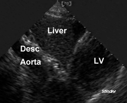
Scanning the inferior vena cava to estimate right atrial pressures
should take into account the possible influence of ascites on caval
diameter changes. (Gastroenterology 1973; 65:294-299)
OBJECTIVE: To investigate the safety, value, and impact of transesophageal echocardiography during liver transplantation. DESIGN: Retrospective. SETTING: University teaching hospital. PARTICIPANTS AND INTERVENTIONS: The medical records of 346 patients and the videotapes of 100 intraoperative transesophageal echocardiography examinations were reviewed. MEASUREMENTS AND MAIN RESULTS: Transesophageal echocardiography was indicated for intraoperative monitoring in 62 patients, 41 of whom had pertinent findings, and for diagnostic purposes in 38 patients, 14 of whom had the expected diagnosis verified. Thirty-one patients had no intraoperative findings. Information that would not have been detected intraoperatively by other means included intracardiac defects, the potential for transpulmonary air passage, valvular regurgitation, the presence or absence of ventricular dysfunction, and embolization occurring at allograft reperfusion. Unanticipated findings during the initial transesophageal echocardiography examination as well as evaluation of intraoperative events resulted in a major impact on patient management in 11% of patients. Preoperatively, 64 patients had a prothrombin time greater than 14 seconds; 56 had a platelet count less than 100,000/mm3; and 23 had esophageal varices, 7 of whom had not had variceal sclerotherapy. Two patients had a complication possibly caused by transesophageal echocardiography (sinus bradycardia and upper gastrointestinal bleeding). No patient experienced documented variceal hemorrhage, esophageal or gastric perforation, and/or oropharyngeal trauma. CONCLUSIONS: It appears that transesophageal echocardiography can be performed safely in patients undergoing liver transplantation, is efficacious in rapidly disclosing new information and monitoring during periods of hemodynamic instability, and may have a significant impact on intraoperative patient management during liver transplantation.
Pulmonary hypertension is now recognized to be a rare association of liver disease and portal hypertension. This report describes the slow resolution of symptomatic pulmonary hypertension in a 33-year-old woman with cirrhosis who underwent isolated liver transplantation. The patient survived the surgery and perioperative period without significant haemodynamic compromise. After liver transplantation, the patient was monitored with regular Doppler echocardiography. By 27 months the pulmonary hypertension had almost completely resolved. This observation is important, as it suggests that patients with severe pulmonary hypertension who survive the perioperative period may have an excellent outcome, although resolution may be slow.
Orthotopic liver transplantation (OLT) in patients with end-stage liver disease is a procedure associated with high cardiac output, low systemic vascular resistance (SVR), coagulopathy and the potential for significant blood loss. A feature of hypertrophic obstructive cardiomyopathy (HOCM) is left ventricular outflow tract obstruction which may be exacerbated by reduced SVR, reduced filling pressures, tachycardia and positive inotropy. We report two cases of OLT in patients with HOCM. Our anaesthetic technique involved the use of halothane and vecuronium and avoidance of drugs causing tachycardia and positive inotropy. Management was aided by intraoperative transoesophageal echocardiography which showed that filling pressures poorly reflected end-diastolic volumes. Volume administration, vasoconstrictors and avoidance of inotropes and chronotropes reduced the outflow tract obstruction which was particularly severe in the reperfusion period.
Orthotopic liver transplantation is an established therapy for end-stage liver disease. This study evaluated the range of cardiovascular abnormalities in patients undergoing evaluation for orthotopic liver transplantation and determined the prognostic implications of abnormal echocardiographic features, including ischemia during dobutamine stress echocardiography, in predicting postoperative cardiac events. Two-dimensional echocardiography was performed in 190 patients for assessment of left ventricular function, valvular pathology, and pulmonary hypertension. Dobutamine stress echocardiography was performed in 165 patients for evaluation of inducible ischemia. Contrast echocardiography for detection of intrapulmonary shunting was performed in 125 patients at rest and in 99 during dobutamine stress. Left ventricular dysfunction, significant valvular regurgitation, and inducible ischemia were identified in <1O% of patients. Pulmonary hypertension, left ventricular hypertrophy and > or = moderate intrapulmonary shunting were present in 12%, 16%, and 26% of patients, respectively. Severe intrapulmonary shunting predicted death prior to transplantation (P=0.01). Of the 71 transplanted patients, major perioperative events included global left ventricular dysfunction in four patients and myocardial infarction in one patient with normal coronary arteries. No preoperative echocardiographic parameters, including ischemia on dobutamine echocardiography, predicted these perioperative events. No cardiac events related to obstructive coronary artery disease occurred in the 154 patients without ischemia on dobutamine stress echocardiography. The majority of patients with end-stage liver disease, including those with alcoholic cirrhosis, have normal cardiac function on two-dimensional echocardiography. Severe intrapulmonary shunting portends a poor prognosis in patients awaiting transplantation. A negative dobutamine stress echocardiogram appears useful in excluding patients at risk for perioperative cardiac events related to obstructive coronary artery disease.
In human liver transplantation, air embolism is seldom encountered after graft reperfusion. Nevertheless, despite adequate flushing and clamping routines, air emboli have been reported in transesophageal echocardiography (TEE) studies performed during the reperfusion phase. We retrospectively investigated whether air in the donor liver -- as observed with pretransplant magnetic resonance imaging (MRI) -- resulted in clinical air embolism or contributed to preservation/reperfusion injury. Clinical air embolism was assessed by intraoperative hemodynamics and end-tidal CO2 monitoring. Preservation/reperfusion injury was assessed in postoperative biochemical measurements. The outcomes were compared between patients receiving livers containing significant intrahepatic air and patients receiving livers without intrahepatic air. Forty-three livers were studied, seven which had major intrahepatic air and ten of which had no evidence of air collections. Twenty-six livers showed minor amounts of air and were excluded from further study. One patient who received a liver that did not contain intrahepatic air had clinical evidence of air embolism. Clinical air embolism did not appear to be associated with the presence of significant intrahepatic air based upon pretransplant MRI. Intrahepatic air did not seem to affect the amount of preservation/reperfusion injury. Our data indicate that air bubbles in the portal and arterial branches are absorbed during reperfusion and that the majority of intrahepatic air is effectively removed by the specific flushing routines.
OBJECTIVE: To review current knowledge about the hepatopulmonary syndrome, including definition and clinical features, methods for diagnosing it, pathophysiologic mechanisms of the associated vascular dilatations, and considerations in treatment, with emphasis on potential reversibility of the syndrome after liver transplantation. DATA SOURCES: The MEDLINE database from January 1986 to December 1993 and bibliographies of selected articles. STUDY SELECTION: Case studies and series reporting results from patients with the hepatopulmonary syndrome were reviewed. Clinical reviews and animal studies relevant to the hepatopulmonary syndrome were examined. DATA EXTRACTION: Outcomes, including survival and the frequency of reversibility of the hepatopulmonary syndrome, were extracted from available clinical reports. DATA SYNTHESIS: Mild hypoxemia is multifactorial and occurs in approximately one third of all patients with chronic liver disease. The hepatopulmonary syndrome is one cause of hypoxemia that may also cause dyspnea, platypnea, and orthopnea. Intrapulmonary vascular dilatations and the resulting right-to-left intrapulmonary shunt are characteristic of the syndrome. Pharmacologic treatment with almitrine bismesylate, somatostatin analog, and indomethacin and treatment with plasmapheresis have been disappointing. The underlying cause and the predictors of reversibility of the hepatopulmonary syndrome remain unknown, but it has recently been shown that such reversibility is possible and that contrast-enhanced echocardiography appears to be the most sensitive diagnostic test for detecting intrapulmonary vascular dilatations. CONCLUSIONS: In the context of persisting uncertainty about the cause and treatment of the hepatopulmonary syndrome, future studies must focus on better understanding the pathophysiology of the hepatopulmonary syndrome, predicting reversibility after liver transplantation, and identifying other treatment options. [References: 123]
The hepatopulmonary syndrome (HPS) is a functional process which is characterized by the triad of liver cirrhosis, intrapulmonary vascular dilatations, and arterial hypoxemia in absence of detectable intrinsic disease of the lung and the heart. The pathophysiological foundation is the presence of a ventilation-perfusion (VA/Q) inequality based on marked vasodilatation of the pulmonary vessels at the precapillary level. Only in critically ill patients limitations of the diffusion of oxygen from the alveolar gas to the capillary blood and intrapulmonary arterio-venous communications will contribute increasingly to the hypoxemia. For diagnosis of HPS arterial blood gases (under condition of room air and 100% oxygen), contrast echocardiography, pulmonary angiography, and multiple inert gas elimination techniques will provide important informations. Regarding recent studies, liver transplantation is the treatment of choice in patients with severe HPS. [References: 24]
Postreperfusion syndrome (PRS) is the most dramatic and acute hemodynamic alteration that occurs in OLT. The aim was to determine heart function by hemodynamic monitoring and transesophageal echocardiography during PRS. 24 nonconsecutive patients were allocated to 2 groups: group A (n = 8), patients with PRS, and group B (n = 16), patients without PRS. Usual hemodynamic data were obtained simultaneously with transesophageal echocardiography recording of the left ventricular imaging in 4 different stages: after induction of anesthesia, 5 min before the end of the anhepatic phase, between 2 and 5 min after reperfusion, and 5 min after graft reperfusion. The hemodynamic and echocardiographic findings during reperfusion were (group A vs. group B patients): mean arterial pressure, 50.0 +/- 15.2 vs. 74.7 +/- 13.9 mmHg (P < 0.01); pulmonary capillary wedge pressure, 12.7 +/- 6.1 vs. 13.9 +/- 5.7 mmHg (NS); left ventricular ejection fraction, 79.6 +/- 9.3 vs. 83.4 +/- 9.4% (NS); left ventricular end diastolic volume index, 35.5 +/- 12.7 vs. 54.7 +/- 21.3 ml/m2 (P < 0.05); and stroke volume index, 27.9 +/- 8.9 vs. 45.5 +/- 15.9 ml/m2 (P < 0.01). There was a mild decrease in left ventricular compliance in group A. There was no alteration in left ventricular function that can justify PRS. The hemodynamic changes during PRS seemed to be caused by an insufficient increase in preload after unclamping.
Severe hypoxemia is an uncommon feature of hepatic cirrhosis. Its major cause are intrapulmonary arteriovenous shunts, both due to direct arteriovenous communications and abnormally dilated pulmonary capillaries. In the present study, a case of cirrhosis associated with severe hypoxemia is reported. Contrast echocardiography showed pulmonary arteriovenous shunts, and low values of mixed venous blood (15% or less were obtained with the method of 100% oxygen breathing). These data suggest that the basic mechanism of hypoxemia, in this case, were capillary dilatations rather than true pulmonary arteriovenous anastomoses. The pathophysiological mechanisms of hypoxemia in cirrhosis are discussed, with emphasis on the present relevance of echocardiography for a full evaluation of these patients, particularly when liver transplantation is contemplated.
A case is reported of a foramen ovale becoming patent during orthotopic liver transplantation (OLT). The patient had a hepatoma secondary to post-hepatitis cirrhosis. Monitoring included transesophageal echocardiography (TEE). A veno-venous shunt between the right femoral, portal and left axillary veins was used so as to maintain the venous return during portal and caval clamping. The patient's haemodynamic state remained quite stable throughout this period, and no vasoactive drug was required. Five min after graft reperfusion, pulmonary arterial pressure increased suddenly (mean PAP: 27 mmHg). TEE revealed paradoxical movements of the atrial septum. Colour coded Doppler ultrasound showed blood flowing from the right to the left atrium through a patent foramen ovale. Fifteen min later, mean PAP decreased (18 mmHg) and TEE no longer showed any flow between the two atria. Several studies have reported transient pulmonary hypertension after unclamping when the donor liver is reperfused. This could induce right ventricular failure, with transient inversion of the atrial pressure gradient, which, in turn, could result in a right-to-left shunt through a patent foramen ovale. TEE can monitor regional and overall left ventricular function as well as the atrial septum. This technique might therefore to be useful for cardiac monitoring during OLT.
In 16 adult patients, continuous intraoperative two-dimensional transesophageal echocardiography (2DTEE) was performed to help elucidate the mechanism of myocardial dysfunction that accompanies liver transplantation. In 4 of the 16 patients "paradoxical" motion of the interventricular septum consistent with right ventricular failure was seen. An additional three of the 16 patients showed right atrial enlargement and right-to-left deviation of the interatrial septum. Two patients showed evidence of paradoxical embolization (one of whom had right ventricular and right atrial enlargement), and a third patient (who had right atrial enlargement) embolized a large right atrial thrombus into the pulmonary circulation. Two-dimensional transesophageal echocardiography demonstrated that isolated right ventricular failure might account for some of the hemodynamic instability seen during liver transplantation. Venous, pulmonary, and paradoxical embolization of air and thrombi documented by transesophageal echocardiography likely contribute to right heart failure.
Aspergillus flavus mural endocarditis was diagnosed after death in a 19-year-old man who had undergone orthotopic liver transplantation 4 months before death. His course was complicated by severe acute graft rejection, which required additional transplants 2 and 4 months, respectively, after the first. Review of the medical literature documented an additional 28 cases of aspergillus endocarditis in patients without prior cardiac surgery. The majority of the patients were immunosuppressed. The most common presenting feature was fever, and embolic phenomena occurred in half of the patients during illness. No blood cultures yielded Aspergillus species. Laboratory findings were nonspecific. The diagnosis was made before death in only seven cases. It was based on histologic examination of either embolectomy tissue (four patients) or skin biopsy tissue (one patient) and on echocardiographic demonstration of vegetations (two patients). Echocardiography failed to show vegetations in five of nine cases tested. Two patients survived. Aspergillus endocarditis should be considered in an immunocompromised host who presents with fever and embolic phenomena, with or without a cardiac murmur, and whose blood cultures are sterile.
The preoperative radiologic imaging workups of 44 pediatric liver transplantation patients were reviewed. Biliary atresia (43%) and metabolic disorders (33%) with end-stage liver disease were the leading indications for pediatric liver transplantation at our institution. The radiologic imaging examinations included chest and skeletal radiography, upper gastrointestinal tract series, abdominal ultrasonography (US), computed tomography, angiography, and contrast echocardiography. Abdominal US (performed in 38 of 44 patients) was the pivotal screening imaging examination; it was invaluable in determining the patency and size of the extrahepatic portal vein and inferior vena cava. Angiography is mandatory if this vascular anatomy is not established with certainty on sonograms or if malrotation is seen on the upper gastrointestinal tract series. Congenital malrotation should be differentiated from small bowel malposition caused by portoenterostomy in patients with biliary atresia. Vascular anomalies, especially absent portal vein and/or inferior vena cava, in patients with biliary atresia and polysplenia syndrome may preclude liver transplantation.
Echocardiographic studies were performed in 73 patients with various types of chronic liver disease. They were 0.5 to 19 years old (mean 5). Thirteen patients underwent follow-up echocardiography 1 to 13 months (mean 6) after liver transplantation. Preoperatively 60 patients (82%) showed evidence of high cardiac output (cardiac index greater than 4 liters/min/m2); these patients manifested increased left ventricular (LV) and left atrial dimensions and a thickened LV posterior wall. Transvenous contrast echocardiographic study confirmed the presence of intrapulmonary arteriovenous shunting in 4 patients. Studies after liver transplantation revealed a reduced LV end-diastolic dimension in 12 patients. Cardiac index was reduced a mean of 35% after transplantation (p less than 0.001). This study suggests that liver transplantation improves common hemodynamic abnormalities in chronic liver disease.
Back to E-chocardiography Home Page.

The contents and links on this page were last verified on November 18, 2012
by Dr. Olga Shindler