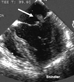
 Mitral valve vegetations in a patient
with systemic lupus erythematosus.
Mitral valve vegetations in a patient
with systemic lupus erythematosus.
Two dimensional echocardiography with doppler examination was performed in 54 patients with systemic lupus erythematosus (SLE). Nine (17%) had significant cardiac involvement (four left ventricular hypertrophy, one moderate pericardial effusion, one severe aortic regurgitation, and three ventricular systolic dysfunction). The authors further studied diastolic function in 45 patients who did not have a major abnormality in echo. SLE was graded as active in 16 patients (SLEDAI > 5) and inactive in 29 patients. Twenty age- and sex- matched subjects acted as controls. The data were compared using one way ANOVA test. Patients with active disease had significant diastolic dysfunction compared to inactive patients and controls as indicated by increased peak A (P < 0.01) and decreased E/A ratio (P < 0.01). There was no linear correlation between disease activity and diastolic dysfunction if SLEDAI was considered as a continuous variable (r=0.29 for E/A). Anticardiolipin antibodies (both IgG and IgM) were elevated in five patients (13 studied). One of them had severe mitral regurgitation, one had trace mitral and aortic regurgitation and one had diastolic dysfunction. The authors conclude that asymptomatic diastolic dysfunction is present in SLE patients.
Kalke S; Balakrishanan C; Mangat G; Mittal G; Kumar N; Joshi VR.
Lupus 1998;7(8):540-4
Prospective two dimensional and Doppler echocardiographic studies were performed in 41 patients to assess the incidence and spectrum of cardiac abnormalities. All patients included in the study fulfilled the 1982 revised criteria of the American Rheumatism Association for classification of SLE. There were 37 women and 4 men with average age of 38 years. Average duration of SLE was 6.5 years (range 6 months to 20 years). Nineteen patients (46.3%) with SLE had cardiac abnormalities. Valvular abnormalities were found in 14 patients (34.1%). Mitral valve abnormalities were the most common findings-in 7 patients (17.1%). There were 6 patients with aortic (14.6%), and 3 patients with tricuspid valve abnormalities (7.3%). One patient had morphological echocardiographic pattern suggesting noninfective verrucous vegetations affecting the tricuspid valve. Pericardial effusion was identified in 5 patients (12.2%). The authors found no correlation between the prevalence of cardiac abnormalities and duration, age and disease activity in SLE patients.
Straburzynska-Migaj E; Leszczynski P; Piszczek I; Cieslinski A;
Mackiewicz S
Pol Merkuriusz Lek 1997 Aug;3(14):76-8
SLE affects most aspects of cardiac function, and recent studies have reported increasing cardiovascular morbidity and mortality. Pathologically, SLE is characterized by a pancarditis involving pericardium, myocardium, endocardium, and coronary arteries. In autopsy series, pericarditis has been found in 43% to 100% (mean 62%, Table I), and myocarditis was found in 8% to 78% (mean 40%, Table II), but both have been underdiagnosed clinically. Libman-Sacks lesions have been noted in 25% to 100% (mean 43%) and infective endocarditis in 1.1% to 4.9% of clinical and autopsy studies. Coronary disease may be due to arteritis, which should be treated with high-dose steroids, or it may be due to atherosclerosis, which is amenable to medical or surgical therapy. Valvular disease has been treated surgically, but with a combined surgical mortality as high as 25%. Aortic insufficiency and mitral regurgitation are the most common valvular problems, although aortic and mitral stenosis have also been reported. Hypertension has been noted in 14% to 69%, and heart failure in 5% to 44%. Evidence for a lupus cardiomyopathy, which may be subclinical, is reviewed. While steroids may ameliorate SLE pancarditis, they have also been associated with hypertension, LV hypertrophy, purulent and constrictive pericarditis, mitral regurgitation, and perhaps accelerated atherosclerosis. It remains to be seen if improved diagnosis and treatment of the cardiovascular manifestations of SLE can enhance survival.
Doherty NE; Siegel RJ
Am Heart J 1985 Dec;110(6):1257-65
Cardiac involvement was noninvasively evaluated in 75 consecutive patients with systemic lupus erythematosus (SLE) by two-dimensional and Doppler echocardiography. In 50/75 patients anticardiolipin antibodies (aCL) were also investigated. Major endocardial damage, characterized by the simultaneous presence of both anatomical and functional valvular involvement (AFVI), was observed in three patients with valvular vegetations and in five patients with combined valvular stenosis and/or regurgitation. Nine patients showed only an anatomic valvular involvement (AVI), expressed by a thickening of one or more valvular leaflets, without echo-Doppler findings of valvular dysfunction. Occurrence of major valvular involvement appears to be correlated with both longer disease duration (9.8 +/- 5.6 yrs in AFVI group vs 5.7 +/- 5.6 yrs in the remaining SLE patients; p < 0.001) and IgG aCL (chi-square = 5.546; p < 0.05). Left ventricular systolic function, evaluated by two-dimensional echocardiographic ejection fraction, was preserved in all patients (EF: 60 +/- 5%). Left ventricular diastolic function, as expressed by echo-Doppler transmitral flow indices of left ventricular filling, was subclinically impaired in 23 patients: only disease duration was significantly longer in these patients (7.7 +/- 5.9 yrs vs 4.9 +/- 4.8 yrs; p < 0.05). Our study demonstrated that cardiac involvement is quite frequent in SLE patients: the disease duration affects both endocardial and myocardial involvement; the anticardiolipin antibodies appear to be related to endocardial but not to myocardial damage.
Migliaresi S, Valentini G, Arnese M, Losardo L, Marone G, Tirri G, Condorelli M. Spectrum of cardiac involvement in systemic lupus erythematosus: echocardiographic, echo-Doppler observations and immunological investigation.Giunta A, Picillo U, Maione S. Acta Cardiol. 1993;48(2):183-97.
Cardiac involvement, evaluated by echo-doppler-cardiography, occurred in 41 of 50 (82%) patients with systemic lupus erythematosus (SLE). Valvular pathology with aortic cusp sclerosis was the most prevalent finding irrespective of age. This finding, suggestive of atherosclerotic heart disease, was supported by increased levels of cholesterol and triglycerides in these patients. There was no significant increase in Lp(a) in the whole patient group, but Lp(a) was raised in patients with proteinuria. Forty percent of the SLE patients had pericarditis. Twelve patients with hypertension and/or mitral regurgitation had increased dimensions of left ventricle, left atrium or interventricular septum while 15 of 50 patients had isolated
increase of these parameters. Localized hypokinesia was found in nine patients. Reduced cardiac index was found in five patients with SLE. There was no association between valvular disease, increased pulmonary artery pressure, and anticardiolipin antibodies.
Rantapää-Dahlqvist S, Neumann-Andersen G, Backman C, Dahlén G, Stegmayr B.
Echocardiographic findings, lipids and lipoprotein(a) in patients with systemic lupus erythematosus.
Clin Rheumatol. 1997 Mar;16(2):140-8.
Mitral valve vegetations in a patient
with systemic lupus erythematosus.
Back to E-chocardiography Home Page.

The contents and links on this page were last verified on August 14, 2012
by Dr. Olga Shindler.