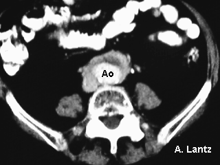
CT scan of the descending aorta with mural thrombus surrounded by soft tissue attenuation encasing the aorta indicating the presence of retroperitoneal fibrosis.
References:
1. Retroperitoneal fibrosis: evaluation by ultrasonography and color
Doppler imaging. Erden A; Aytaç S; Cumhur T; Yurdakul M;
Calikoglu U. Urol Int, 55(2):111-4 1995
Delay in diagnosis is common in retroperitoneal fibrosis because of
the non-specific clinical presentation. Ultrasonography combined with
color Doppler imaging is a rapid and practical method in the early
diagnosis and during follow-up. The authors describe 3 cases with
retroperitoneal fibrosis, emphasizing the ultrasonographic and color
Doppler features of the disease.
2. Marfan syndrome and retroperitoneal fibrosis. Phillips S; Evans C. Am J Roentgenol, 167(6):1596-7 1996 Dec
Back to E-chocardiography Home Page.
e-mail:shindler@umdnj.edu