Classic M-mode appearance of mitral stenosis: decreased EF slope, leaflet thickening, anterior motion of the posterior mitral leaflet.
Hockey stick appearance of the anterior mitral leaflet in mitral stenosis.
Color flow outline of a decreased mitral valve orifice area in the short axis view.
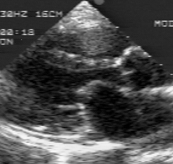
Diastolic color Doppler flow convergence in the apical four chamber view of mitral stenosis.
Chest films showing left atrial enlargement. There is straightening of the left heart border (above) and splaying of the carina (below).
The auscultatory hallmarks of mitral stenosis include a loud first heart sound, a diastolic opening snap, and a diastolic rumble with presystolic accentuation. The patient should be in the left lateral decubitus position for detection of the diastolic rumble. The bell of the stethoscope should be applied lightly at the apex at the point of maximal impulse.
If the diastolic rumble remains inaudible - one should listen at the apex again, but this time while the patient is actually turning from the supine to the left lateral decubitus position. The reason for this is that in some patients the diastolic rumble disappears quickly after turning and would be missed if one were to wait for the patient to complete the turn before applying the stethoscope.
MP3 audio file of auscultatory features of mitral stenosis. This is a large file (more than 800,000 bytes). It shows the increased intensity of the first heart sound. The second heart sound is normal. It is followed by an opening snap. The intensity of the opening snap was manipulated electronically to change in loudness from beat to beat so the listener would recognize it more readily. Normally, the opening snap does not vary from beat to beat.
Four types of mitral stenosis were noted: commisural, cuspal, chordal and combined.
There were two types of commisural stenosis:
1. The commisure retains its normal depth but is abnormal by virtue of increase in thickness and loss of pliability.
2. The depth of the commisure is increased because of fusion of the valvular leaflets in the vicinity of the commisure. The newly formed component of the commisure is continuous with the original commisure.
In each of these two subgroups the anterolateral and posteromedial commisures may be affected equally or only one commisure may be involved.
Cuspal type of mitral stenosis is conversion of the mitral leaflets or cusps into stiff rigid and often leathery membranes. The anterior leaflet is usually involved to a greater extent.
Chordal type of mitral stenosis results from thickened,shortened and frequently fused chordae. Because each leaflet is connected to opposing chordae, chordal shortening causes reduced flexibility of the leaflets even when the leaflet structure is unaffected.
Dobutamine Stress in Mitral Stenosis
J Am Coll Cardiol. 2002 Nov 20;40(10):1809-15.
Is the mitral valve area flow-dependent in mitral stenosis? A dobutamine stress echocardiographic study.
Mohan JC, Patel AR, Passey R, Gupta D, Kumar M, Arora R, Pandian NG.
Department of Cardiology, G. B. Pant Hospital, New Delhi, India.
OBJECTIVES: The purpose of this study was to compare the effect of changes in flow rate on the mitral valve area (MVA) derived from two-dimensional echocardiographic planimetry and Doppler pressure half-time (PHT) methods in patients with mitral stenosis (MS). BACKGROUND: Dobutamine stress echocardiography has been proposed as a means of assessing the severity of MS. However, data regarding the effect of an increase in flow rate on MVA are limited. If MVA is indeed flow-dependent, this has important implications for the assessment of the severity of MS, particularly in the setting of reduced cardiac output (CO). METHODS: Dobutamine echocardiography was performed in 57 patients with isolated MS who were in sinus rhythm. The MVA was determined by planimetry and Doppler PHT methods. RESULTS: Cardiac output increased by > or =50% in 27 patients (group I) and by <50% in 30 patients (group II). In group I, the MVA by planimetry increased by only 10.6 +/- 2% and the MVA by PHT increased by 21.9 +/- 4.8%. These changes were similar to those observed in group II (10.7 +/- 3% and 14.8 +/- 4%, respectively; p = NS), despite a much smaller increase in CO. A clinically important change (from the severe to mild category) occurred in only one patient when using the PHT method and in none by planimetry. CONCLUSIONS: Changes in flow rate result in small but clinically insignificant changes in echocardiographic MVA measurement. These methods provide an accurate assessment of MS severity in a majority of patients, independent of changes in flow rate.
Am J Cardiol. 1997 Nov 15;80(10):1374-7.
Comparison of exercise and dobutamine stress echocardiography in assessing mitral stenosis.
Hecker SL, Zabalgoitia M, Ashline P, Oneschuk L, O'Rourke RA, Herrera CJ.
Division of Cardiology, Illinois Masonic Medical Center, Chicago 60657, USA.
Dobutamine elicited similar hemodynamic response to exercise in 20 consecutive patients with mitral stenosis, and significantly altered management in 6 of them (30%). Dobutamine stress echocardiography is a safe and feasible alternative to exercise in patients with mitral stenosis of mild-to-moderate severity and ambiguous symptoms.
Am Heart J. 1986 Feb;111(2):312-6.
The usefulness of dobutamine in the assessment of the severity of mitral stenosis.
Hwang MH, Pacold I, Piao ZE, Engelmeier R, Scanlon PJ, Loeb HS.
Patients with mitral stenosis often require supine exercise in order to increase their heart rate and cardiac output to assess the severity of their valvular obstruction during cardiac catheterization. We substituted dobutamine for exercise in 14 patients with suspected mitral stenosis. The dobutamine infusion was started at 5 micrograms/kg/min and was increased to 10, 15, and 20 micrograms/kg/min every 3 minutes as tolerated. The heart rate increased from 84 +/- 4 to 123 +/- 7 bpm (p less than 0.001), the cardiac index increased from 2.4 +/- 0.2 to 3.4 +/- 0.2 L/min/m2 (p less than 0.001), and the mean pulmonary artery pressure increased from 27 +/- 3 to 30 +/- 2 mm Hg (p less than 0.02). The pulmonary wedge pressure of 19 +/- 2 mm Hg and the mitral valve index of 0.8 +/- 0.1 cm2/m2 remained unchanged, but the left ventricular end-diastolic pressure decreased from 11 +/- 2 to 6 +/- 2 mm Hg (p less than 0.02). The hemodynamic response during the infusion of dobutamine identified a subgroup of patients with more severe mitral stenosis. Thus, the administration of dobutamine is useful in the evaluation of the severity of mitral valve obstruction during catheterization.
Back to E-chocardiography Home Page.
e-mail:shindler@umdnj.edu
The contents and links on this page were last verified on July 15, 2002.