Cardiac Myxomas
Intracardiac myxomas are most commonly found in the left atrium. The tumor is
typically pedunculated. The most common point of attachment is at the
atrial septum in the region of the fossa ovalis. Once the diagnosis is
made, the tumor is removed surgically. Follow up echocardiograms are
performed to rule out recurrence. The references below reflect the
different opinions on rate of recurrence.
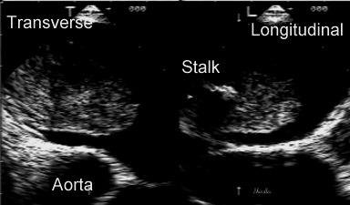
The biplane image below displays the stalk and the
tissue characteristics of the myxoma.
Selected References
-
King TW. On simple vascular growths in the left auricle of the heart.
Lancet 1845; 2:428-9.
(First description of a myxoma).
-
Effert S. Domanig E. Diagnostik intraaurikularer tumoren und grosser thromben
mit dem ultraschall-echoverfahren. Dtsch Med Wochenschr 1959;
84:6-8.
(First echocardiographic description of a myxoma).
-
Lane GE, Kapples EJ, Thompson RC, Grinton SF, Finck SJ. Quiescent
left atrial myxoma. Am Heart J 1994;
-
Pochis WT, Wingo MW, Cinquegrani MP, Sagar KB. Echocardiographic
demonstration of rapid growth of a left atrial myxoma. Am Heart J
1991; 122:1781-4.
-
Malekzadeh S, Roberts WC. Growth rate of left atrial myxoma. Am J Cardiol
1989; 64:1075-6.
-
Roudaut R, Gosse P, Dallocchio M. Rapid growth of a left atrial
myxoma shown by echocardiography. Br Heart J 1987; 58:413-6.
(Rate of growth using echocardiography)
-
Waller DA, Ettles DF, Saunders NR, Williams G. Recurrent cardiac
myxoma:
the surgical implications of two distinct groups of patients. Thorac
Cardiovasc Surg 1989, 37:226-30.
-
Gray IR, Williams WG. Recurring cardiac myxoma. Br Heart J 1985,
53:645-9. (Rate of recurrence - the reason for follow-up echocardiographic
studies after myxoma resection)
Links to Genetics, Case Reports, Images, Pathology
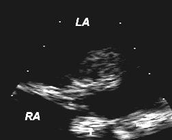
 Transesophageal image demonstrating the typical pedunculated attachment
to the interatrial septum of a left atrial myxoma.
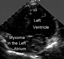
 Transthoracic apical view of a left atrial myxoma.
 Biplane transesophageal view showing simultaneous transverse and
longitudinal planes of a left atrial myxoma. The stalk is visible
in the longitudinal plane.
Back to E-chocardiography Home Page.

The contents and links on this page were last verified on
July 3, 2006.
 Transesophageal image demonstrating the typical pedunculated attachment
to the interatrial septum of a left atrial myxoma.
 Transesophageal image demonstrating the typical pedunculated attachment
to the interatrial septum of a left atrial myxoma.
 Transthoracic apical view of a left atrial myxoma.
 Biplane transesophageal view showing simultaneous transverse and
longitudinal planes of a left atrial myxoma. The stalk is visible
in the longitudinal plane.
