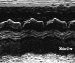
M-mode of the pulmonic valve showing early systolic closure and an absent A wave in a patient with severe pulmonary hypertension.
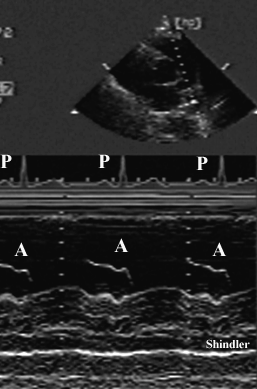
Normal pulmonic A wave in a patient with normal pulmonary artery pressures (shown for comparison).
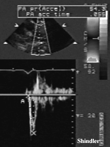
Pulsed wave Doppler measurement of pulmonary artery systolic acceleration in a patient with pulmonary hypertension.
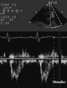
Pulsed wave Doppler in the pulmonary artery showing rapid early systolic acceleration and mid-systolic slowing.
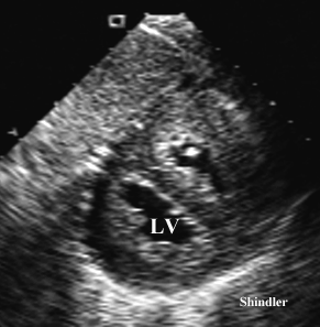
Transgastric transesophageal view, showing right ventricular hypertrophy and abnormal contour of the interventricular septum - resulting in a D shaped left ventricle.
J Am Coll Cardiol. 1987 Mar;9(3):549-54.
Comparison of three Doppler ultrasound methods in the prediction of pulmonary artery pressure.
Chan KL, Currie PJ, Seward JB, Hagler DJ, Mair DD, Tajik AJ.
Pulmonary artery pressure was noninvasively estimated by three Doppler echocardiographic methods in 50 consecutive patients undergoing cardiac catheterization. First, a systolic transtricuspid gradient was calculated from Doppler-detected tricuspid regurg itation; clinical jugular venous pressure or a fixed value of 14 mm Hg was added to yield systolic pulmonary artery pressure. Second, acceleration time from pulmonary flow analysis was used in a regression equation to derive mean pulmonary artery pressure . Third, right ventricular isovolumic relaxation time was calculated from Doppler-determined pulmonary valve closure and tricuspid valve opening; systolic pulmonary artery pressure was then derived from a nomogram. In 48 patients (96%) at least one of the methods could be employed. A tricuspid pressure gradient, obtained in 36 patients (72%), provided reliable prediction of systolic pulmonary artery pressure. The prediction was superior when 14 mm Hg rather than estimated jugular venous pressure was used to account for right atrial pressure. In 44 patients (88%), pulmonary flow was analyzed. Prediction of mean pulmonary artery pressure was unsatisfactory (r = 0.65) but improved (r = 0.85) when only patients with a heart rate between 60 and 100 beats/min w ere considered. The effect of correcting pulmonary flow indexes for heart rate was examined by correlating different flow indexes before and after correction for heart rate. There was a good correlation between corrected acceleration time and either systo lic (r = -0.85) or mean (r = -0.83) pulmonary artery pressure. Because of a high incidence of arrhythmia, right ventricular relaxation time could be determined in only 11 patients (22%). Noninvasive prediction of pulmonary artery pressure is feasible in m ost patients.
Am J Cardiol. 1986 Aug 1;58(3):352-6.
Factors affecting use of the Doppler-determined time from flow onset to maximal pulmonary artery velocity for measurement of pulmonary artery pressure in children.
Serwer GA, Cougle AG, Eckerd JM, Armstrong BE.
Measurement of the time from onset to maximal or peak velocity (TPV) of pulmonary artery (PA) flow has been proposed as a noninvasive means of determining PA pressure. The effects of age, heart rate, increased PA pressure and flow, pulmonary valve obstruc tion and altered PA vascular resistance on this measurement were evaluated. In 84 children, aged 1 day to 18 years, TPV was measured using continuous-wave Doppler echocardiography. The children were separated into 3 groups. Group I (n = 33) consisted of c hildren with no cardiovascular abnormalities. Group II (n = 33) consisted of children with a variety of cardiovascular diseases producing varying PA pressures and flows. Group III (n = 18) consisted of children who had valvular pulmonic stenosis with PA t o right ventricular gradients greater than 40 mm Hg. Doppler studies of group II and III patients were performed in conjunction with measurement of PA pressures and flows at the time of cardiac catheterization. In group I TPV showed a significant negative linear correlation with heart rate (r = -0.86, p less than 0.001). The ratio of observed TPV to predicted TPV (TPVN) determined using the regression equation for TPV vs heart rate or TPV/TPVN was heart rate- and age-independent (p greater than 0.1) and r anged from 0.81 to 1.31 (mean 1.005). In group II TPV/TPVN was inversely related to the natural log of the PA pressures (systolic, r = -0.91; mean, r = -0.87; diastolic, r = -0.82; all p less than 0.01), whether pressure elevation was due to increased flo w, resistance or left atrial hypertension.
Am J Cardiol. 1990 Aug 15;66(4):493-6.
Noninvasive estimation of right atrial pressure from the inspiratory collapse of the inferior vena cava.
Kircher BJ, Himelman RB, Schiller NB.
University of California, San Francisco, Division of Cardiology, John Henry Mills Echocardiography Laboratory.
To evaluate a simple noninvasive means of estimating right atrial (RA) pressure, the respiratory motion of the inferior vena cava (IVC) was analyzed by 2-dimensional echocardiography in 83 patients. Expiratory and inspiratory IVC diameters and percent col lapse (caval index) were measured in subcostal views within 2 cm of the right atrium. Parameters were correlated with RA pressure by flotation catheter within 24 hours of the echocardiogram (38 were simultaneous). Correlations between RA pressure (range 0 to 28 mm Hg), expiratory and inspiratory diameters and caval index were 0.48, 0.71 and 0.75, respectively. Of 48 patients with caval indexes less than 50%, 41 (89%) had RA pressure greater than or equal to 10 mm Hg (mean +/- standard deviation, 15 +/- 6) , while 30 of 35 patients (86%) with caval indexes greater than or equal to 50% had RA pressure less than 10 mm Hg (mean 6 +/- 5). Sensitivity and specificity for discrimination of RA pressure greater than or equal to or less than 10 mm Hg were maximized at the 50% level of collapse. Thus, IVC respiratory collapse on echocardiography is easily imaged and can be used to estimate RA pressure. A caval index greater than or equal to 50% indicates RA pressure less than 10 mm Hg, and caval indexes less than 50% indicate RA pressure greater than or equal to 10 Hg.
J Am Soc Echocardiogr. 1992 Nov-Dec;5(6):613-9.
Does inferior vena cava size predict right atrial pressures in patients receiving mechanical ventilation?
Jue J, Chung W, Schiller NB.
Department of Medicine, University of California, San Francisco.
The inferior vena cava diameter and its respiratory response are used to estimate right atrial pressures in spontaneously breathing patients but its value in patients receiving mechanical ventilation is unvalidated. Forty-nine patients undergoing mechanic al ventilation were prospectively evaluated in the intensive or coronary care units with two-dimensional echocardiography of the inferior vena cava and simultaneous measurements of mean right atrial pressures by central venous or pulmonary artery catheter . Correlation between inferior vena cava diameter at expiration and mean right atrial pressure was only 0.58. The correlation between inspiratory change in inferior vena cava diameter and mean right atrial pressure was poor (r = 0.13). Despite these corre lations, an inferior vena cava diameter of < or = 12 mm predicted a right atrial pressure of 10 mm Hg or less 100% of the time, but sensitivity was only 25%. An inferior vena cava diameter > 12 mm had no predictive value for right atrial pressure.
Circulation. 1986 Sep;74(3):484-92.
Continuous-wave Doppler echocardiographic detection of pulmonary regurgitation and its application to noninvasive estimation of pulmonary artery pressure.
Masuyama T, Kodama K, Kitabatake A, Sato H, Nanto S, Inoue M.
Continuous-wave Doppler echocardiography was used to estimate pulmonary artery pressures by measuring pulmonary regurgitant flow velocity in 21 patients with pulmonary hypertension (mean pulmonary artery pressure greater than or equal to 20 mm Hg) and 24 patients without pulmonary hypertension. The pulmonary regurgitant flow velocity patterns, characterized by a rapid rise in flow velocity immediately after closure of the pulmonary valve and a gradual deceleration until the next pulmonary valve opening, w ere successfully obtained in 18 of the 21 patients with pulmonary hypertension and in 13 of the 24 patients without pulmonary hypertension. As pulmonary artery pressure increased, pulmonary regurgitant flow velocity became higher; the pulmonary artery-to- right ventricular pressure gradient in diastole (PG) was estimated from the pulmonary regurgitant flow velocity (V) by means of the simplified Bernoulli equation (PG = 4V2). The Doppler-determined pressure gradient at end-diastole correlated well with the catheter measurement of the pressure gradient at end-diastole (r = .94, SEE = 3 mm Hg) and with pulmonary artery end-diastolic pressure (r = .92, SEE = 4 mm Hg). The peak of Doppler-determined pressure gradient during diastole correlated well with mean p ulmonary artery pressure (r = .92, SEE = 5 mm Hg). Thus continuous-wave Doppler echocardiography was useful for noninvasive estimation of pulmonary artery pressures.
Circulation. 1983 Aug;68(2):302-9.
Noninvasive evaluation of pulmonary hypertension by a pulsed Doppler technique.
Kitabatake A, Inoue M, Asao M, Masuyama T, Tanouchi J, Morita T, Mishima M, Uematsu M, Shimazu T, Hori M, Abe H.
We used a pulsed Doppler technique to examine the flow velocity pattern in the right ventricular outflow tract in 33 adults. In the patients with normal pulmonary artery pressure (mean pressure less than 20 mm Hg, 16 patients), ejection flow reached a pea k level at midsystole (137 +/- 24 msec, mean +/- SD), producing a domelike contour of the flow velocity pattern during systole. In contrast, the flow velocity pattern in patients with pulmonary hypertension (mean pressure greater than or equal to 20 mm Hg , 17 patients) was demonstrated to accelerate rapidly and to reach a peak level sooner (97 +/- 20 msec, p less than .01); in 10 of the pulmonary hypertensive patients a secondary slower rise in flow velocity was observed during a deceleration, resulting i n the midsystolic notching. The time to peak flow (acceleration time, AcT) and right ventricular ejection time (RVET) were measured from the flow velocity pattern. Either AcT or AcT/RVET decreased with increase in mean pulmonary artery pressure, and a ver y high correlation (r = -.90) was found between AcT/RVET and log10 (mean pulmonary artery pressure). The use of this technique permitted the noninvasive estimation of the pulmonary artery pressure.
Back to E-chocardiography Home Page.

The contents and links on this page were last verified on December 2, 2004.