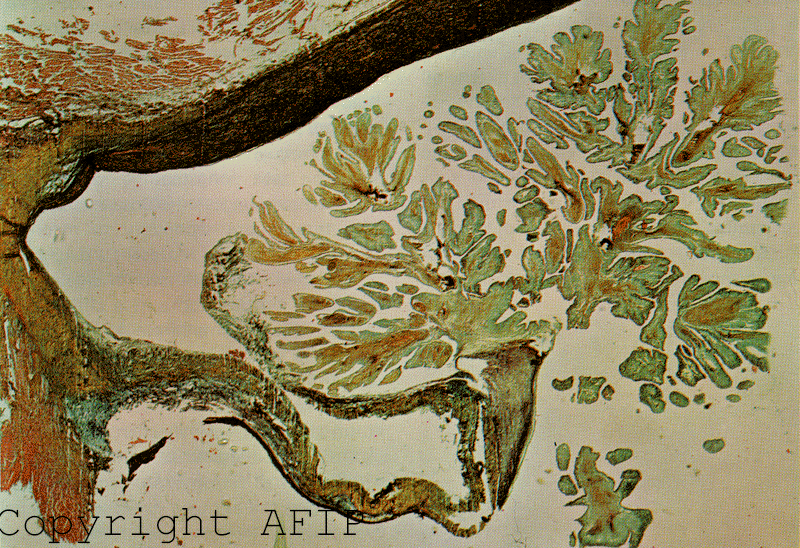
 Papillary fibroelastoma of the tricuspid valve.
Copyright Armed Forces Institute of Pathology.
Papillary fibroelastoma of the tricuspid valve.
Copyright Armed Forces Institute of Pathology.
Papillary fibroelastoma of the tricuspid valve.
Copyright Armed Forces Institute of Pathology.
Mitral valve repair for anterior leaflet papillary fibroelastoma: two case
descriptions and a literature review
Di Mattia DG; Assaghi A; Mangini A; Ravagnan S; Bonetto S; Fundaro P
Eur J Cardiothorac Surg 1999 Jan;15(1):103-7
Cardiac papillary fibroelastomas are rare cardiac tumors and have been considered a `benign' incidental finding that may have significant clinical manifestations. In this paper we report two cases of mitral valve fibroelastoma: one was discovered by chance with transthoracic echocardiography in a young healthy man, the other was an intraoperative incidental finding in a middle aged man with a recent history of acute myocardial infarction. The mitral valve was repaired in both cases after excising the tumor. The patients did well and remain asymptomatic. A literature review was compiled which comprises previous case reports of 34 patients with mitral valve papillary fibroelastomas. Most were asymptomatic, but when symptoms occurred, they could be disabling, such as stroke, cardiac heart failure, myocardial infarction, and sudden death. Papillary fibroelastoma is amenable to simple surgical excision or in addition to mitral valve repair or replacement. Recurrence has not been reported.
Cardiac papillary fibroelastoma: a rare cause of ischemic stroke in the young
Giannesini C; Kubis N; N'Guyen A; Wassef M; Mikol J; Woimant F
Cerebrovasc Dis 1999 Jan-Feb; 9(1): 45-9
Among etiologies of stroke in young adults, primary cardiac tumors are very rare. We report the case of a 37-year-old woman who was admitted for an ischemic stroke in the right middle cerebral artery region. Etiologic investigations revealed, after transthoracic and transesophageal echocardiography, an aortic valve tumor. Treatment was first medical with anticoagulation, then surgical. Histological examination showed a papillary fibroelastoma. After a review of the literature, the possible mechanisms of the ischemic event are discussed and lead to the conclusion that this tumor must be surgically excised, even if asymptomatic, because of recurrent ischemic complications responsible for myocardial infarction, stroke and sudden death.
Cardiac valve papillary fibroelastoma: surgical excision for revealed or potential
embolization
Grinda JM; Couetil JP; Chauvaud S; D'Attellis N; Berrebi A; Fabiani JN; Deloche
A; Carpentier A
J Thorac Cardiovascu Surg 1999 Jan;117(1):106-10
OBJECTIVE: We have reviewed the case histories of 4 patients who underwent operations between September 1994 and November 1997 at Broussais Hospital for cardiac valvular papillary fibroelastoma. METHODS: Diagnosis was strongly suggested by echocardiography. Tumor locations were mitral (1), tricuspid (1), and aortic (2). Indications for operation were previous stroke for the mitral tumor, prophylaxis for the tricuspid tumor, syncopal episodes for the first aortic tumor, and transient ischemic attack and mesenteric ischemia for the second aortic tumor. RESULTS: Surgical excision with a conservative, valve-sparing approach was performed in all cases. For the first aortic tumor, aortic valve reconstruction was achieved with part of a cryopreserved aortic homograft cusp. Intraoperative transesophageal echocardiography showed no evidence of valvular regurgitation after excision in all cases. All patients had uneventful postoperative recoveries. No evidence of regurgitation or recurrence was seen on echocardiography at follow-up. CONCLUSIONS: Despite their histologically benign aspect, cardiac papillary fibroelastomas should be excised because of potential embolic complications. A conservative, valve-sparing approach is recommended, however, because of the absence of recurrence after total excision.
A papillary fibroelastoma of the left ventricle in the presence of a mitral valve
prosthesis. A case report and review of the literature.
Roman Herrera L; Pech Escalante CM; Martinez Enriquez A; Hernanez Chavez
VG; Melendez Lopez C; Alva Espinosa C
Arch Inst Cardiol Mex 1998 May-June; 68(3): 232-8
Papillary fibroelastoma is a rare, benign cardiac tumor. Before echocardiogram came into existence, it was diagnosed only by necropsy or incidentally at surgery. This kind of tumor may appear on the endocardial surface or in any of the valves. Although it is usually small in size, it is associated to embolic phenomena, thoracic pain and sudden death. This report presents the first case of papillary fibroelastoma in the presence of a mechanical valvular prosthesis in mitral position. In a patient 55 years old, presenting inactive rheumatic heart disease. The tumor was detected by means of transthoracic and transesophageal echocardiogram.
Calcified papillary fibroelastoma of the tricuspid valve
Paelinck B; Vermeersch P; Kockx M
Acta Cardiol 1998; 53(3): 165-7
Papillary fibroelastoma (PFE) is an infrequently and often incidentally encountered benign tumor found on the endocardium. We describe the case of a patient with a small, mobile, calcified mass on the tricuspid valve incidentally seen during radioscopy for cardiac catheterization. Echocardiography revealed a rounded, bulky shaped and calcified tumor. At histopathology a PFE with extensive dystrophic calcifications was found. To our knowledge this is the second case report of calcified PFE.
Papillary fibroelastoma. A rare etiology of strokes in young patients
Bailbe M; Coisne D; Babin P; Corbi P; Menu P; Rosier MP; Couderq C; Pouget
Abadie JF; Gil R; Neau JP
Rev Med Interne 1998 Feb; 19(2): 119-22
BACKGROUND: The papillary fibroelastomas are cardiac lesions, which typically occur on the cardiac valves, but rarely on the endocardium. The coincidence of these benign primitive tumors varies from 0.002 to 0.33% and increases with advancing age. METHODS: We report two cases of stroke, one in a 31 year old man and the other in a 48 year old woman, both admitted to the same stroke center. RESULTS: The diagnostic studies were normal in these two patients, except for the echocardiography. The first showed an echogenic mass on the mitral valve on transthoracic echocardiography (TTE), confirmed by the transesophageal echocardiography (TEE). The second demonstrated a mass on the sigmoid aortic valve on TEE, but the TTE was normal. For these two patients, a surgical excision was carried out and pathologic examination concluded to a papillary fibroelastoma. After surgery, no recurrence was observed. CONCLUSIONS: The papillary fibroelastomas are usually asymptomatic and easily detected by TEE. However, it can be revealed by stroke, myocardial infarction and lower limbs ischemia. These cardiac tumors should be surgically removed, since their complete excision remains the only means of avoiding a recurrence of embolism.
Papillary fibroelastoma of the heart (giant Lambl excrescence). Clinical-
anatomical study on 10 surgically treated patients.
Loire R; Pinede L; Donsbeck AV; Nighoghossian N; Perinetti M
Presse Med 1998 Apr 25; 27(16): 753-7
OBJECTIVES: The growing number of reports of surgery for papillary fibroelastomas of the heart led us to evaluate the diagnostic potential of ultrasonography in patients with cerebral or coronary signs and to assess the efficacy of anticoagulant therapy in preventing recurrent cerebral ischemia and disease progression after resection. PATIENTS AND METHODS: Ten cases of echographically diagnosed fibroelastoma of the heart treated by surgery were analyzed together with cases reported in the literature. RESULTS: Transesophageal echography has been shown to be the superior method. Surgical resection has given good results and the postoperative course is always excellent. Recurrent embolism occurred in two of our cases despite well- conducted anticoagulation. DISCUSSION: Surgical resection should be performed as early as possible because anticoagulation does not appear to sufficiently protect against embolic events, particularly cerebral events.
Clinical expression of papillary fibroelastoma.
Gully C; Benghanem MM; Motebassem R; Sagan C
Arch Mal Coeur Vaiss 1998 Jun; 91(6): 777-82
The authors review the literature of the clinical features of papillary fibroelastomas in the light of a new case. These benign tumors of the endocardium may be distinguished from Lambl's vegetations by their site and size. Some workers suggest that they correspond to giant Lambl's vegetation and could be a form of "aging" of the valvular endocardium. Nevertheless, Lambl's vegetations are always present after 10 years of age but the papillary fibroelastoma is rarely detected by echocardiography and there have been few case reports. They are essentially cardiac valve tumours (73% of valvular tumours) and may give rise to serious clinical symptoms, sudden death by migration or coronary obstruction, systemic embolism, especially from left heart lesions. However, they can be situated at any point of the endocardium. The diagnosis of a valvular or an endocardial tumour is based on echocardiography which, though not always accurate, gives a better etiological diagnosis. In cases of symptomatic tumour, surgery (usually simple ablation) is indicated with a low operative risk and cure of symptoms. Tumours discovered by chance pose very difficult problems of management and may lead to diagnostic or preventive surgery.
Echocardiographic detection of pulmonary valve papillary fibroelastoma
Bhagwandien NS; Shah N; Costello JM Jr; Gilbert CL
J Cardiovasc Surg (Torino) 1998 Jun; 39(3): 351-4
Papillary fibroelastomas of the heart are rare lesions usually discovered at autopsy or incidentally at surgery. Although these lesions are benign and generally asymptomatic, they can cause valvular dysfunction or embolize to vital structures. In this case report, we describe a pulmonary valve papillary fibroelastoma detected by echocardiography in an adult. Most of the 12 cases of pulmonary valve papillary fibroelastoma reported in the literature were discovered incidentally at autopsy or during surgery. To our knowledge, this is the first reported case of pulmonary valve papillary fibroelastoma detected by echocardiography. Rest imaging before exercise echocardiography for evaluation of atypical chest pain in a 42 year old white female demonstrated a mass on the pulmonary valve. The mass was further characterized by transesophageal echocardiography and excised during open heart surgery. Pulmonary valve papillary fibroelastoma was diagnosed histopathologically. This case illustrates the additional diagnostic value of comprehensive 2D imaging in the rest phase before doing exercise echocardiography.
Evolution of a papillary fibroelastoma
Malik MF; Sagar K; Wynsen JC; Kenny D
J Am Soc Echocardiogr 1998 Jan;11(1):92-4
Papillary fibroelastoma is a rare primary tumor of the heart usually found
incidentally at autopsy. Little is known about the natural history of this tumor, but
an aggressive surgical approach is recommended because of the high incidence
of embolization. We describe a patient whose tumor was found during
transthoracic echocardiography and who had had a normal echo 10 years
previously. This finding suggests that papillary fibroelastoma may be an
acquired rather than a congenital lesion.
Cardiac papillary fibroelastoma. Different forms of the clinical presentation
Caballero J; Calle G; Arana R; Sancho M
Rev Esp Cardiol 1997 Nov; 50(11): 815-7
Papillary fibroelastoma is an uncommon cardiac tumor rarely diagnosed during
life. Although most fibroelastomas are incidental findings at autopsy, a few
cases have been associated with cardiac symptoms that include angina, arterial
embolism and sudden death. We report the case of two patients, a 35 year old
male with an acute myocardial infarction and ventricular fibrillation and a 53 year
old asymptomatic female, with cardiac masses first detected by transthoracic
echocardiography. A more detailed morphological study was provided by
transesophageal echocardiography. After cardiac surgery, the anatomical study
demonstrated that both tumors were papillary fibroelastomas. The literature
concerning papillary fibroelastoma is reviewed.
Left ventricular papillary fibroelastoma: two-dimensional echocardiographic
detection and surgical resection
Jobic Y; Etienne Y; Quintin-Roue I; Dewilde J; Cornec P; Gilard M; Le Bras Y;
Barra JA; Loire R; Boschat J; et al
J Am Soc Echocardiogr 1996 Sep-Oct; 8(5PT 1): 756-8
We report a patient with a papillary fibroelastoma arising from the left ventricular
posterior wall. The tumor was detected incidentally during echocardiography
undertaken to evaluate aortic stenosis. Possible complication from tumor
embolization was avoided by surgical resection during aortic valve replacement.
Incidental detection of an aortic valve papillary fibroelastoma by
echocardiography in an asymptomatic patient presenting with hypertension.
Evans AJ; Butany J; Omran AS; David TE
Can J Cardiolo 1997 Oct; 13(10): 905-8
Papillary fibroelastomas are rare, frond-like tumours of uncertain etiology seen
on cardiac valves, uncommonly found antemortem. They carry a significant risk
of embolization, making their detection and excision during life an important
issue. A case of an aortic valve papillary fibroelastoma is described, which was
found at echocardiography in a patient being assessed for previously
unrecognized, severe hypertension.
Echocardiographic features of papillary fibroelastoma and their consequences
and management
Yee HC; Nwosu JE; Lii AD; Velasco M; Millman A
Am J Cardiol 1997 Sep 15; 80(6): 811-4
Thirty-five percent of patients (5 of 15) were diagnosed with ischemic stroke from
left-sided papillary fibroelastomas by diagnosis of exclusion, whereas 40% of
patients (6 of 15) did not have ischemic stroke.
Cardiac papillary fibroelastoma excision combined with reconstructive surgery
Tkebuchava T; von Segeser LK; Gallino A; Dirsch O; Turina MI
Jpn Heart J 1997 May; 38(3): 457-62
Three cases of cardiac papillary fibroelastomas are described. Two-dimensional
echocardiography detected the tumors in the mitral valve, the cordae tendinae
and in the apex of the left ventricle-a unique location. The tumor excisions
were combined with bypass operation, mitral valve reconstruction, repair of
cordae tendinae and Maze-procedure. The three patients are doing well after
surgery.
Papillary fibroelastoma: echocardiographic characteristics for diagnosis and
pathologic correlation
Klarich KW; Enriquez-Sarano M; Gura GM; Edwards WD; Tajik AJ; Seward JB
J Am Coll Cardiol 1997 Sep; 30 (3): 784-90
OBJECTIVES: We sought to determine the clinical and echocardiographic
characteristics of papillary fibroelastoma (PFE). BACKGROUND: PFE is a
rarely encountered cardiac tumor about which relatively little is known.
METHODS: Institutional records were reviewed for the years 1980 to 1995 for
patients with pathologic or echocardiographic diagnosis of PFE. Group 1
included 17 patients with the pathologic diagnosis of PFE who also underwent
echocardiography. Echocardiographic features of PFE were established in
group 1. Group 2 included 37 patients with only echocardiographic evidence of
PFE. RESULTS: In group 1, 7 (41.2%) of 17 patients had symptoms related to
PFE. Neurologic events occurred in 5 (29.4%) of 17 patients. All patients had
the tumor surgically removed. During follow-up, no new embolic events
occurred. Echocardiographic characteristics of PFE included a small tumor
(12.1 +/- 6.5 x 9.0 +/- 4.3 mm), usually pedunculated (14 [94%] of 17 patients)
and mobile, with a homogeneous speckled pattern and a characteristic stippling
along the edges. PFEs were most common on valvular surfaces (12 [60%] of 20
PFEs) but were not uncommon on other endocardial surfaces (8 [40%] of 20
PFEs). The tumor did not cause valvular dysfunction. In group 2, 16 (43%0 of
37 patients were asymptomatic. Five patients (13.5%) had a previous
neurologic event. During follow-up (mean 31 months, range 1 to 77), nine
neurologic events occurred. CONCLUSIONS: PFEs are associated with
embolism, can be diagnosed with echocardiography, are often an incidental
clinical finding and do not cause valvular dysfunction.
Papillary fibroelastoma of the tricuspid valve
Bentley MJ; Mullen JC
Can J Cardiol 1997 Aug;13(8):773-4
A 50 year old woman with a long standing history of palpitations was found by
echocardiography to have a 1.5 cm mass on the atrial surface of the anterior
leaflet of the tricuspid valve. Surgical excision included a portion of surrounding
leaflet tissue. The tumor was a papillary fibroelastoma. She remained
asymptomatic with no recurrence in the follow-up.
Echocardiographic diagnosis of papillary fibroelastoma of the mitral and tricuspid
valve apparatus
Lund GK; Schroder S; Koschyk DH; Nienaber CA
Clin Cardiol 1997 Feb;20(2):175-7
Papillary fibroelastomas are rare and normally benign cardiac tumors typically
attached to cardiac valves. This report describes two patients who were
evaluated for intermittent dyspnea in one case and for the source of cerebral
embolism in the other. In both patients transthoracic echocardiography revealed
a pedunculated mobile mass adjacent to an atrioventricular valve, suggestive of
papillary fibroelastoma. Postoperative histology was confirmatory of papillary
fibroelastoma with a typical hyalinized hypocellular stroma covered by a single
layer of endocardial cells.
Visualization of ventricular fibroelastoma with a video-assisted thoracoscope
Espada R; Talwaker NG; Wilcox G; Kleiman NS; Verani MS
Ann Thorac Surg 1997 Jan;63(1):221-3
Left ventricular papillary fibroelastomas are associated with a high risk of
cerebral embolization. Two-dimensional echocardiography and intraoperative
transesophageal echocardiography are helpful in diagnosing tumors, planning a
surgical approach, and achieving adequate excision. A video-assisted
thoracoscope via the left atrium was used to visualize a left ventricular papillary
fibroelastoma. Thoracoscopic visualization facilitated excision of a mass within
the chordae tendinae between the anterolateral papillary muscle and the left
ventricular wall. Video-assisted thoracoscopy greatly facilitates
exposure/excision of deeper intracavitary left ventricular masses.
Papillary fibroelastoma of the aortic valve in a patient with an acute myocardial
infarction
Pasteuning WH; Zijnen P; van der Aa MA; Peters JH
J Am Soc Echocardiogr 1996 Nov-Dec;9(6):897-900
We describe a patient with myocardial infarction in whom a tumor near the aortic
valve was identified by routine transthoracic echocardiography.
Transesophageal echocardiography proved to be particularly useful in identifying
the attachment of the tumor to the aortic valve and enabled a surgical approach
through the ascending aorta. On microscopic examination, the tumor appeared
to be a papillary fibroelastoma.
Cardiac papillary fibroelastoma
Ni Y; von Segesser LK; Dirsch O; Schneider J; Jenni R; Turina M
Thorac Cardiovasc Surg 1996 Oct;44(5):257-60
Papillary fibroelastomas are rare and benign heart tumors. We present two
cases with these lesions. A young female patient with cerebral infarction was
operated to resect the tumor on the mitral valve and the valve was successfully
repaired. Another male patient had a history of bradycardia. On examination,
coronary stenosis and a tumor in the left ventricle was found. Tumor excision
combined with aorto-coronary artery bypass grafting was performed.
Echocardiography proved to be highly effective to diagnose these tumors.
Because of the potential cerebral and coronary embolization, these tumors
should be excised. Surgical results are good.
A case report of mitral valve papillary fibroelastoma leading to embolic stroke
Shirota K; Yano Y; Hayase S; Ogawa K; Fujita K
Kyobu Geka 1996 Jul;49(7):571-4
A 39-year-old man presented with acute onset of left arm and left side face
weakness, and mild expressive aphasia. He was referred for two-dimensional
echocardiography, which demonstrated a 13-mm diameter pedunculated, mobile,
echodense mass attached to the anterior leaflet of the mitral valve. At operation,
the tumor was a 10 x 10 x 15 mm, rounded, and yellow-whitish mass with a short
stalk arising from rough zone of the anterior leaflet of the mitral valve. The
surface appeared to be sean anemone. The mitral valve with this tumor was
excised and replaced with a 29 mm St. Jude Medical mechanical valve. The
pathological findings are typical of a papillary fibroelastoma.
A case report of papillary fibroelastoma of the aortic valve
Hirota J; Akiyama K; Ookado A; Takiguchi M; Oosawa S; Hashimoto A
Nippon Kyobu Geka Gakkai Zasshi 1996 May;44(5):705-8
A 69-year-old woman referred to our hospital with an aortic valve tumor. She
had shown signs of chronic heart failure due to atrial fibrillation and hypertension
for 4 years. There was no history of thromboembolism such as stroke and
myocardial infarction, unaccountable fever, weight loss, and systemic symptoms.
With two-dimensional echocardiography, a cardiac valve tumor was detected
during a routine examination for heart failure. Echocardiographic findings
showed a homogenous mass with a diameter of approximately 1.5 cm, fixed
directly to the noncoronary aortic valve cusp. During the operation, a papillary
neoplasm, 1.5 by 1 cm, was observed at the midportion of the left ventricular side
of noncoronary cusp without a peduncle. The tumor was excised together with
all cusps. A 21 mm SJM aortic valve was implanted in position, and thereafter
she remained free from symptoms. Histopathological examination of the tumor
revealed benign papillary fibroelastoma. Two-dimensional echocardiography
was utilized for a diagnosis of the aortic papillary fibroelastoma.
Echocardiographic evaluation of papillary fibroelastoma: a case report and
review of the literature.
Hicks KA; Kovach JA; Frishberg DP; Wiley TM
J Am Soc Echocardiogr 1996 May-Jun; 9(3):353-60
Papillary fibroelastomas comprise approximately 7.9% of benign primary cardiac
tumors. Although papillary fibroelastomas were at first discovered incidentally at
autopsy or during heart surgery, these tumors are increasingly being identified by
echocardiography. This article reviews those papillary fibroelastomas detected
by transthoracic or transesophageal echocardiography and discusses the
echocardiographic features of these tumors, associated symptoms, and
management. Echocardiography is important in influencing management
decisions regarding excision, valve replacement, and valve repair.
Papillary fibroelastoma of the mitral valve in a 3-year-old child: case report
De Menezes IC; Fragata J; Martins FM
Pediatr Cardiol 1996 May-Jun; 17(3): 194-5
A 3.5-year-old boy with stroke secondary to embolization of a primary cardiac
tumor is presented. The diagnosis was made by two-dimensional
echocardiography and confirmed intraoperatively. A papillary fibroelastoma was
identified histologically. It is a rare condition and a diagnostic challenge, as
patients are asymptomatic before embolization. In view of the severe
consequences, prompt surgery is recommended.
Papillary fibroelastoma of the tricuspid valve in association with an atrial septal
defect: report of a case
Watanabe T; Hosoda Y; Kikuchi N; Kawai S
Surg Today 1996;26(10):831-3
Although papillary fibroelastoma is rare, it is the most common primary tumor of
the heart valves. We describe herein the case of a 64 year old woman
scheduled to undergo atrial septal defect (ASD) repair, in whom a papillary
fibroelastoma of the tricuspid valve was diagnosed by transesophageal
echocardiography (TEE). Surgical resection of the papillary fibroelastoma at
the time of ASD repair prevented the fatal embolization sometimes associated
with this lesion. Thus, intraoperative TEE played an important role in identifying
the location of the tumor and its anatomic attachment, and in assessing the
adequacy of surgical treatment.
Cardiac papillary fibroelastoma: a treatable cause of transient ischemic attack
and ischemic stroke detected by transesophageal echocardiography
Brown RD Jr; Khandheria BK; Edwards WD
Mayo Clin Proc 1995; 70:863-8
Transesophageal echocardiography (TEE) is used frequently in patients with
cerebrovascular ischemia. On TEE, a typical appearance of a cardiac
fibroelastoma is that of pedunculated, mobile mass attached to a leaflet of a
valve. Surgical excision of the lesion may lead to resolution of the symptoms
and prevent further cerebrovascular ischemic events; valve replacement is
seldom necessary. Herein we describe three patients with cerebral or ocular
ischemia in whom histologic study confirmed a cardiac papillary fibroelastoma
after initial detection by TEE. Cardiac papillary fibroelastomas should be
considered in the differential diagnosis of transient ischemic attach and stroke,
even in cases of recurrent events in the same vascular distribution. Although
the use of echocardiography in the evaluation of stroke and transient ischemic
attack is controversial, TEE must be considered in patients in whom the cause of
cerebrovascular ischemia echocardiography, even if the patient's cardiac history
and the findings on physical examination are normal.
Excision of papillary fibroelastoma arising from the septal leaflet of the tricuspid
valve
Lee CC; Celik C; Lajos Tz
J Card Surg 1995 Sep;10(5):589-91
A case of a papillary fibroelastoma (PFE) arising from the tricuspid valve was
reported. It was incidentally detected by two-dimensional transthoracic
echocardiography. Prior to 1977, these tumors were exclusively found at
postmortem examination. This is only the fourth reported case of a tricuspid
valve PFE found by echocardiography, treated by excision, and with tricuspid
valvuloplasty preserving the native valve.
Multiplane transesophageal echocardiography detection of a papillary
fibroelastoma of the aortic valve causing myocardial infarction
Grote J; Mugge A; Schfers HJ; Daniel WG; Lichtlen PR
Eur Heart J 1995 Mar; 16(3): 426-9
Primary aortic valve tumours are rare. A patient is reported with a papillary
fibroelastoma attached to the edge of the right coronary aortic cusp. This
tumour was diagnosed by multiplane transesophageal echocardiography after
the patient experienced an acute inferolateral myocardial infarction. Multiplane
transesophageal echocardiography was useful to visualize and identify the
precise point of attachment of the tumour, enabling cardiac surgeons to plan
aortic valve repair rather than replacement.
Surgical treatment of right atrial papillary fibroelastoma, originated from the
Eustachian valve-a case report
Shigemitsu O; Hadama T; Mori Y; Miyamoto S; Sako H; Uchida Y;
Nippon Kyobu Geka Gakkai Zasshi 1995 Mar;43(3):403-6
Papillary fibroelastoma is one of the most common benign primary cardiac tumor
after myxoma. However, it is rare to originate from Eustachian valve. A 44-
year-old woman was pointed out the right atrial tumor during admission for
vasospastic angina. On two-dimensional echocardiogram, the tumor was found
in the right atrium, and easily moved to the right ventricle. At operation, the
tumor originated from Eustachian valve, and was resected together with the
valve by means of extracorporeal circulation. The size of tumor was 57 mm in
length and the histological diagnosis was papillary fibroelastoma. The
postoperative course was uneventful.
Cardiac papillary fibroelastoma
Shahian DM; Labib SB; Chang G
Ann Thorac Surg 1995 Feb; 59(2): 538-41
Papillary fibroelastomas are rare cardiac tumors, but they are the most common
primary tumor of the heart valves. These lesions occur on any of the valves or
endothelial surfaces of the heart and may be detected by echocardiography,
cardiac catheterization, during open heart operation for other conditions, or at
autopsy. Because of their potential for cerebral and coronary embolization, even
small papillary fibroelastomas should be excised.
Papillary fibroelastoma of the mitral valve. A rare cause of embolic events.
Colucci V; Alberti A; Bonacina E; Gordini V
Tex Heart Inst J 1995;22(4):3227-31
A 66-year-old woman was admitted to our department with an 11-month history
of multiple transient ischemic attacks and strokes. A 2 dimensional
echocardiographic study revealed an intracardiac tumor attached both to the
chordae and to the anterolateral papillary muscle of the mitral valve. The
patient underwent excision of the tumor, which necessitated concomitant mitral
valve replacement. S she remains free of symptoms 1 year postoperatively, with
no echocardiographic evidence of recurrence of the tumor. To date, 19 cases of
surgically treated papillary fibroelastomas of the mitral valve have been reported
in the English-language literature. We add the description of our case to
emphasize the importance of this tumor as an identifiable and curable cause of
cerebral and coronary embolization. The frequent occurrence of cardiac valve
tumors suggests the use of 2-dimensional echocardiography in patients who are
experiencing transient ischemic attacks or strokes, as well as in those who
sustain a myocardial infarction despite normal coronary arteries at angiography.
When papillary fibroelastoma is diagnosed, surgical treatment must be
considered because of the high risk of embolization.
Pseudopapillary fibroelastoma of the mitral valve
Madu E; Myles J; Fraker TD Jr
J Natl Med Assoc 1995 Jan;87(1):68-70
Papillary fibroelastomas are well-recognized benign cardiac neoplasms. They
are primarily asymptomatic, but occasionally are associated with neurologic and
cardiac symptoms. Pseudopapillary fibroelastomas presenting with usual
clinical and echocardiographic manifestations of papillary fibroelastoma but
lacking characteristic histologic features have not been described previously.
This article describes a 42-year-old, previously healthy female admitted with
sudden hemiparesis and dysarthria. Symptoms completely resolved within 4
days. Extensive investigations revealed no etiology except for a pedunculated
mitral valve mass with echocardiographic appearance suggestive of papillary
fibroelastoma. Histologic staining, however, failed to reveal characteristic
features of papillary fibroelastoma.
Papillary fibroelastoma of the mitral valve associated with rheumatic mitral
stenosis
Bedi HS; Sharma VK; Mishra M; Kasliwal RR; Trehan N
Eur J Cardiothorac Surg 1996;9(1):54-5
Papillary fibroelastoma of the mitral valve diagnosed and treated in life is
extremely rare. There have been eight cases documented so far. We report
the first case of a mitral valve papillary fibroelastoma associated with severe
rheumatic mitral stenosis and tricuspid regurgitation with stenosis. The tumor
arose from the posteromedial papillary muscle of the mitral valve. The mitral
valve was replaced after excising the valve with the tumor and the tricuspid valve
was repaired. The patient did well and remains asymptomatic.
Embolizing fibroelastoma of the aortic valve
Ragni T; Grande AM; Cappuccio G; Arbustini E; Grasso M; Tramarin R; Vigano
M
Cardiovasc Surg 1994 Oct;2(5):639-41
Papillary fibroelastomas are very rare cardiac tumors that can present with
embolization of coronary and peripheral arteries and sudden death. The
diagnosis can be made by two-dimensional or transesophageal
echocardiography. A 53-year-old man with no aortic valve papillary
fibroelastoma who presented with several transient ischemic attacks is reported.
Incidental finding of papillary fibroelastoma on the atrial septum
Nakao T; Hollinger I; Attai L; Oka Y
Cardiovasc Surg 1994 Jun; 2(3): 423-4
Papillary fibroelastoma is rare but one of the most common benign primary
cardiac tumors after myxoma. This lesion may be associated with embolization,
angina and sudden death. The incidental finding of a small pedunculated
papillary fibroelastoma arising from the atrial septum detected by
transesophageal two-dimensional echocardiography (TEE) in a patient
undergoing coronary artery bypass grafting is reported. The advantage of TEE
in diagnosing intracardiac tumors is also described.
Emboligenic mitral papillary fibroelastoma and positive antiphospholipid
antibodies
Roldan Torres I; Salvador Sanz A; Mora Llabata V; Marti Llinares S; Chirivella
Gonzalez A; Vera Sempere F; Hernandez Martinez M; Campayo Ibanez A;
Algarra Vidal FJ
Rev Esp Cardiol 1994 Apr; 47(4): 255-7
Cardiac papillary fibroelastoma has been associated to high levels of
antiphospholipid antibodies, either primary or in the context of systemic lupus
erythematosus. We present the case of a young female with several episodes
of peripheral emboli. Two-dimensional echocardiography demonstrated a tumor
on the anterior mitral leaflet. The mass was resected and histologically showed
a papillary architecture covered by hyperplasic endocardial cells on a layer of
connective tissue and a central core of collagen and elastic fibers. The
immunologic study demonstrated high titers of anticardiolipin antibodies,
complement consumption and positive antinuclear antibodies. The patient keep
high anticardiolipin antibodies titers at follow-up but embolization has not
recurred and has no symptoms.
Transesophageal echocardiographic localization of an aortic valve papillary
fibroelastoma during routine coronary artery bypass surgery
Autz L; Krieger KH; Yao FS
J Heart Valve Dis 1993 Nov; 2(6): 662-4
Papillary fibroelastomas are rare, primary cardiac tumors most often diagnosed
incidentally at autopsy. These tumors have been associated with embolism,
neurologic injury, coronary ischemia and sudden death. We report a case of
clinical detection of any asymptomatic aortic valve papillary fibroelastoma by
transesophageal echocardiography. The mass was an incidental finding during
routine coronary artery bypass surgery. This finding dictated a change in the
operative approach to include resection of the aortic valve mass in addition to
coronary revascularization.
Papillary fibroelastoma of the mitral valve
Shapira OM; Williamson WA: Duga JM
Cardiovascu Surg 1993 Oct; 1(5): 599-601
A case report of a 73-year-old woman with mitral regurgitation secondary to
papillary fibroelastoma and prolapse of the mitral valve is described. The tumor
was excised, and the valve repaired with Duran annuloplasty ring. The
clinicopathologic features and the surgical management of this rare tumor are
reviewed.
A symptomatic papillary fibroelastoma of the left ventricle removed with the aid of
transesophageal echocardiography
Schuetz WH; Welz A; Heymer B
Thorac Cardiovasc Surg 1993 Aug; 41(4): 258-60
A papillary fibroelastoma, causing amaurosis fugax and paresis of the right arm,
was detected by echocardiography as a free floating mass in the left-ventricular
outflow tract. Based on the exact localization of the tumor by intraoperative
transesophageal echocardiography the initial indication for ventriculotomy was
disregarded and an atraumatic transvalvular approach was chosen.
Transesophageal echocardiography in the detection and surgical management of
a papillary fibroelastoma of the mitral valve causing partial mitral valve
obstruction
Thomas MR; Jayakrishnan AG; Desai J; Monaghan MJ; Jewitt DE
J Am Soc Echocardiogr 1993 Jan-Feb; 6(1): 83-6
Primary mitral valve tumors are rare. We describe the transesophageal
appearances of a papillary fibroelastoma (Lambl's giant excrescence) of the
anterior mitral valve leaflet causing partial mitral valve obstruction.
Transesophageal echocardiography proved particularly useful in identifying the
limited attachment of the tumor to the anterior mitral valve leaflet and excluding
its attachment of the tumor to the anterior mitral valve leaflet and excluding its
attachment to the interatrial septum. These features helped to exclude the
possibility of the tumor being a left atrial myxoma, the primary differential
diagnosis of the lesion. Transesophageal echocardiography enabled the
planned surgical option to be mitral valve repair and also allowed intraoperative
monitoring to assess the results of the surgical repair.
Papillary fibroelastoma in the left ventricular outflow tract
Uchida S; Obayashi N; Yamanari H; Matsubara K; Saito D; Haraoka S
Heart Vessels 1992; 7(3): 164-7
We report a case of a papillary fibroelastoma originating from the left ventricular
endocardium in the outflow tract which was discovered by echocardiography in
an asymptomatic patient. Two echocardiographic features were observed: (1)
the tumor surface was smooth, and characteristic papillary formation was not
detected; and (2) the outline of the mass was clearly defined as a dense echo,
with the central, radiolucent, portion surrounded by a highly refractive linear echo
at the level of the maximum diameter of the mass. The excised tumor was
covered with a gelatinous substance that masked multiple papillae on the
surface, but its echolucent center could not be explained by the pathology of the
tumor which was solid centrally. Our case indicates that a papillary
fibroelastoma may sometimes show echocardiographic findings similar to those
of a myxoma, although other investigators have not noted the smooth surface
and the echolucent center makes it indistinguishable from a myxoma. Thus, in
some cases, it is difficult to distinguish papillary fibroelastoma from myxoma by
echocardiography.
Young patient with left brain infarct and transient right-sided hemiparesis in
cardiac papillary fibroelastoma
Kroll W; Nellessen U; Hofig M; Luttges J; Sievers H; Simon R
Z Kardiol 1991 Mar; 80(3): 234-6
This is a case report of a 25 year old female with recurrent episodes of transient
cerebral ischemia accompanied by transient hemiparesis. Cardiac ultrasound
examination revealed a small left-ventricular tumor located between the chordae
tendineae. The echocardiographic finding was subsequently confirmed by
surgery. The entire tumor was removed and the mitral valve remained intact.
Due to the histological and immunohistochemical findings a papillary
fibroelastoma was diagnosed. The early and late postoperative course was
without event.
Tricuspid valve papillary fibroelastoma: echocardiographic characterization
Wolfe JT 3d; Finck SJ; Stafford RE: Persellin ST
Ann Thorac Surg 1991 Jan; 51(1):116-8
We report a tricuspid valve papillary fibroelastoma initially detected by
transthoracic two-dimensional echocardiography and subsequently characterized
by transesophageal two-dimensional echocardiography. The mass was excised
during open heart operation, and the diagnosis was verified grossly and
histopathologically. Transesophageal echocardiography usually provides
images far superior to those from transthoracic echocardiography and may be a
useful adjunct for intraoperative localization of intracardiac tumors for
excision.
Papillary fibroelastoma of the aortic valve: primary echocardiography diagnosis in
an asymptomatic patient
Rieckmann C; Liebau G; Hellberg K; Seeliger H
Z Kardiol 1990 Mar; 79(3): 208-10
A rare case of a cardiac tumor located between the right coronary ostium and
cusp of the aortic valve is reported. Diagnosis was confirmed by two-
dimensional echocardiography. Histologic evaluation revealed a papillary
fibroelastoma.
Mitral valve fibroelastoma
Gorton ME; Soltanzadeh H
Ann Thorac Surg 1989 Apr; 47(4): 605-7
Papillary fibroelastomas are rare benign tumors of the heart with predisposition
for cardiac valvular involvement that were traditionally incidental findings at
autopsy. Echocardiography now is allowing clinicians to diagnose these tumors
in living patients. We reviewed the literature and, to our knowledge, are
reporting the fifth documented case of a mitral valve papillary fibroelastoma in a
living patient. Our patient was seen with bilateral transient ischemic attacks and
was found to have a mitral valve tumor by two-dimensional echocardiography
and cardiac catheterization. The tumor involved the entire mitral valve and
subvalvular structures. The patient's valve was replaced with a bioprosthesis,
and she remains free from symptoms.
Left ventricular papillary fibroelastoma with coronary embolization
Mazzucco A; Bortolotti U; Thiene G; Dan M; Stritoni P; Scutari M; Stellin G
Eur J Cardiothorac Surg 1989; 3(5): 471-3
We describe a patient who survived an acute myocardial infarction caused by
coronary embolization from a left ventricular papillary fibroelastoma. The
tumour, which was detected by 2-D echocardiography, was successfully excised.
Intracardiac papillary fibroelastoma: an occult cause of embolic neurologic deficit
McFadeen PM; Lacy JR
Ann Thorac Surg 1987 Jun; 43(6): 667-9
We report the case of a young woman with a transient ischemic attack secondary
to a rare intracardiac tumor, a papillary fibroelastoma. The tumor was identified
by two-dimensional echocardiography and was treated surgically. The
intracardiac papillary fibroelastoma as an uncommon source of occult cerebral
emboli is discussed.
Two-dimensional echocardiographic detection and diagnostic features of
tricuspid papillary fibroelastoma
Frumin H; O'Donnell L; Kerin NZ; Levine F; Nathan LE Jr; Klein SP
J Am Coll Cardiol 1983 Nov; 2(5): 1016-8
Cardiac papillary fibroelastomas are rare and benign primary tumors in the
cardiac valves or occasionally the mural endocardium. Before 1977, these
tumors were diagnosed exclusively at postmortem examination. Over the last
few years, a handful of cases have been diagnosed in vivo by echocardiography.
In this report, we describe the first tricuspid valve papillary fibroelastoma
detected by echocardiography in an adult. Clinical and echocardiographic
features are discussed.
Papillary fibroelastoma of the heart. Report of six cases.
Almagro UA; Perry LS; Choi H; Pintar K
Arch Pathol Lab Med 1982 Jul; 106(7): 318-21
Papillary fibroelastoma is a rare cardiac lesion, mostly encountered as an
incidental finding at the time of autopsy but occasionally seen during life.
Although this condition generally is asymptomatic, a few cases had been
associated with clinical problems, such as embolic phenomena, angina pectoris,
outflow tract obstruction, and sudden death. We studied six examples of this
lesion, five occurring on the cardiac valves and one on the left ventricular
septum. In one, the lesion was detected by echocardiography, which represents
to our knowledge only the third published case in which the lesion was visualized
by this diagnostic procedure and only the fourth in which the lesion was seen
during life. We believe that echocardiography is useful in evaluating whether a
case, by virtue of its size or location, carries a significant risk of causing clinical
problems and therefore requires treatment.
Back to E-chocardiography Home Page.

The contents and links on this page were last verified on November 12, 2001.