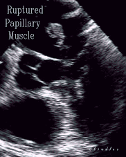

Transesophageal echocardiogram demonstrating papillary muscle rupture following inferior myocardial infarction.
Sanders RJ, Neubuerger KT, Ravin A. Rupture of papillary muscles: occurrence of rupture of the posterior muscle in posterior myocardial infarction. Dis Chest. 1957;31:316-321.
Esthes EH, Dalton FM, Entman ML, Dixon HB, Hackel DB. The anatomy and blood supply of the papillary muscles of the left ventricle. Am Heart J. 1966;71:356-362.
De Busk RF, Harrison DC. The clinical spectrum of papillary muscle disease. N Engl J Med. 1969;281:1458-1462.
Vlodaver Z, Edwards JE. Rupture of ventricular septum or papillary muscle complicating myocardial infarction. Circulation. 1977;55:815-822.
Wei JY, Hutchins GM, Bulkley BM. Papillary muscle rupture in fatal acute myocardial infarction: a potentially treatable form of cardiogenic shock. Ann Intern Med. 1979;90:149-152.
Nishimura RA, Schaff HV, Shub C, Gersh BJ, Edwards WD, Tajik AJ. Papillary muscle rupture complicating acute myocardial infarction: analysis of 17 patients. Am J Cardiol. 1983;51:373-377.
Glock Y, Herreros J, Chaffai M, Carrier D, Sanchez R, Arcas R, Cerene A, Puel P. Le remplacement valvulaire associé au pontage aorto-coronaire dans l'insufficiance mitrale ischémique. Arch Mal Coeur. 1985;78:869-875.
Barbour DJ, Roberts WC. Rupture of a left ventricular papillary muscle during acute myocardial infarction: analysis of 22 necropsy patients. J Am Coll Cardiol. 1986;8:558-565.
Coma-Canella I, Gamallo C, Martinez Onsurbe P, Jadraque LM. Anatomic findings in acute papillary muscle necrosis. Am Heart J. 1989;118:1188-1192.
J Am Coll Cardiol. 1988 Jan;11(1):59-65.
Contrast echocardiography during coronary arteriography in humans: perfusion and anatomic studies.
Feinstein SB, Lang RM, Dick C, Neumann A, Al-Sadir J, Chua KG, Carroll J, Feldman T, Borow KM.
Section of Cardiology, University of Chicago Medical Center, Illinois 60637.
In humans, the physiologic relation between myocardial blood flow and epicardial coronary artery anatomy remains poorly defined. With the recent development of sonicated microbubble contrast agents, it is now possible to use contrast echocardiography to assess myocardial perfusion and to correlate blood flow with angiographically identified coronary artery anatomy. The purpose of the current study was to determine myocardial perfusion patterns in patients without significant coronary artery disease. The results may be used as a reference to analyze myocardial blood flow in patients with coronary artery disease. Sonicated meglumine sodium diatrizoate solution (Renografin-76), which contains microbubbles measuring 4.5 +/- 2.8 micrograms in diameter by laser analysis, was used as the echocardiographic contrast agent during elective coronary arterriography in 14 patients without significant coronary artery disease. Patients received intracoronary injections of 1.5 to 2 ml of sonicated Renografin-76 without complications. Perfusion characteristics were studied by visual assessment of the two-dimensional echocardiographic images obtained after individual injections. In patients found to be free of significant coronary artery disease by arteriography, the left coronary system always supplied the anteroseptal, anterior, anterolateral and posterior regions of the left ventricle at the mid-papillary, cross-sectional level. The right coronary artery system perfused the inferior and inferoseptal regions in 89% of the patients identified with a right dominant system. The anterolateral papillary muscle was perfused from the left coronary system in all cases. The posteromedial papillary muscle was perfused from the left coronary system in 58% of the patients and from the right system in 42% of the patients.
Am J Cardiol. 1996 Oct 15;78(8):955-8.
Left ventricular papillary muscle perfusion assessed with myocardial contrast echocardiography.
Lim YJ, Masuyama T, Nanto S, Mishima M, Kodama K, Hori M.
Cardiology Division, Kawachi General Hospital, Higashi Osaka.
This myocardial contrast echocardiographic study shows that left ventricular posteromedial papillary muscle is supplied by either the right or left coronary artery in most subjects, but may be supplied by both coronary arteries. The posteromedial papillary muscle and its adjacent area may be supplied by a different coronary artery.
Back to E-chocardiography Home Page.
e-mail:shindler@umdnj.edu The contents and links on this page were last verified on April 14, 2005.