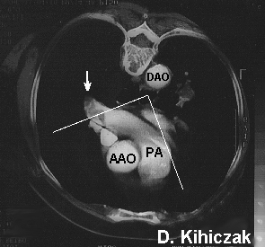
CT scan of the chest showing a thrombus in the right pulmonary artery (arrow). Echocardiography from the esophagus is capable of imaging this area in some patients as shown by the superimposed scanning sector lines.
Hunter JJ; Johnson KR; Karagianes TG; Dittrich HC.
Detection of massive pulmonary embolus-in-transit
by transesophageal echocardiography.
Chest 1991 Nov;100(5):1210-4
Unexplained shock and acute pulmonary hypertension
were evaluated with echocardiography. Standard
transthoracic echocardiography failed to identify
a large embolism-in-transit that was easily
visualized by transesophageal imaging.
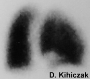
Perfusion scan showing a defect in the right lower lobe.
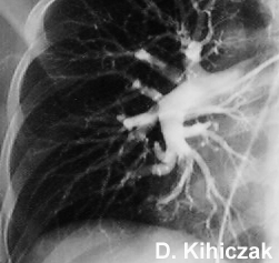
Pulmonary angiogram showing abrupt tapering of a right lower lobe vessel.
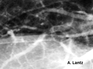
Luminal pulmonary arterial filling defects.
Back to E-chocardiography Home Page.
e-mail:shindler@umdnj.edu