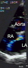
 Short axis parasternal view of a narrow color flow jet in the right atrium.
These images illustrate the capability of transthoracic studies to confirm the presence of a patent foramen ovale in some patients. Intravenous echo contrast may also be useful. Transesophageal echocardiography provides better resolution of the interatrial septum.
Short axis parasternal view of a narrow color flow jet in the right atrium.
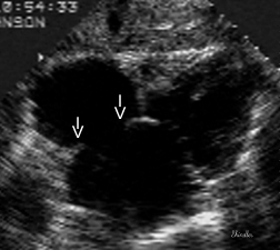 Subcostal image in the same patient that raises the question of atrial
septal defect. The membrane of the fossa ovalis is thinner than the rest
of the atrial septal wall - resulting in echocardiographic dropout that
mimics a non restrictive atrial septal defect.
Subcostal image in the same patient that raises the question of atrial
septal defect. The membrane of the fossa ovalis is thinner than the rest
of the atrial septal wall - resulting in echocardiographic dropout that
mimics a non restrictive atrial septal defect.
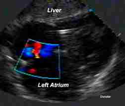
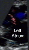 Color flow (red) in the same patient confirms a small left to right shunt
(rather than a large atrial septal defect). Contrast echocardiography is
also useful. The right cardiac chambers were not dilated.
Color flow (red) in the same patient confirms a small left to right shunt
(rather than a large atrial septal defect). Contrast echocardiography is
also useful. The right cardiac chambers were not dilated.
Transesophageal image of a patent foramen ovale at the University of Chicago Echocardiography Laboratory.
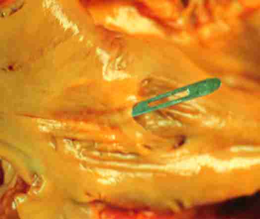 Pathological specimen illustrating the left atrial
appearance of the membrane of the fossa ovalis. The probe is lifting the
thin membrane. Under normal conditions the left atrial pressure is greater
than the right atrial pressure, keeping the foramen closed.
Pathological specimen illustrating the left atrial
appearance of the membrane of the fossa ovalis. The probe is lifting the
thin membrane. Under normal conditions the left atrial pressure is greater
than the right atrial pressure, keeping the foramen closed.
 Subcostal view of a patent foramen ovale in an
elderly patient with marked biatrial enlargement.
Subcostal view of a patent foramen ovale in an
elderly patient with marked biatrial enlargement.
Back to E-chocardiography Home Page.
![]()
The contents and links on this page were last verified on March 20, 2013
by Dr. Olga Shindler