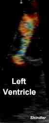
The proximal isovelocity surface area (PISA) of a regurgitant color flow jet can be useful in the estimation of valvular insufficiency. PISA is based on the hemodynamic principles of flow through a small circular orifice in a flat plate. There is flow acceleration just proximal to the orifice. The flow converges on the orifice in hemispheric layers of equal velocity. The surface area of a hemisphere is calculated by the formula: two pi radius squared.
Color flow Doppler can be used to calculate the area of the hemisphere. This area in centimeters squared is then multiplied times the aliasing velocity in centimeters per second yielding cubic centimeters per second, which provides the volume of regurgitant blood flow.
The principal theoretic limitation is that the usual regurgitant orifice is neither circular nor flat.
Furthermore, it is possible to calculate the effective regurgitant orifice area (ERO) by dividing regurgitant flow volume by the velocity of the mitral insufficiency jet: ERO = Flow / Velocity
This is based on conservation of energy. The flow velocity "V" in a vessel will vary inversely with the area of the orifice. Since flow equals velocity times the orifice area, multiplying both sides of the equation: " Flow = Velocity multiplied by the Area (V X A)" by 1/V yields: " Area = Flow / Velocity ".
J Am Coll Cardiol. 1994 Feb;23(2):443-51.
Effective regurgitant orifice area: a noninvasive Doppler development of an old hemodynamic concept.
Enriquez-Sarano M, Seward JB, Bailey KR, Tajik AJ.
Division of Cardiovascular Diseases and Internal Medicine, Mayo Clinic, Rochester, Minnesota 55905.
OBJECTIVES. The purpose of this study was to determine the feasibility, relation to other methods and significance of the effective regurgitant orifice area measurement. BACKGROUND. Assessment of the severity of valvular regurgitation (effective regurgitant orifice area) has not been implemented in clinical practice but can be made by Doppler echocardiography. METHODS. Effective regurgitant orifice area was calculated by Doppler echocardiography as the ratio of regurgitant volume/regurgitant jet time-velocity integral and compared with color flow Doppler mapping, angiography, surgical classification, regurgitant fraction and variables of volume overload. RESULTS. In 210 consecutive patients examined prospectively, feasibility improved from the early to the late experience (65% to 95%). Effective regurgitant orifice area was 28 +/- 23 mm2 (mean +/- SD) for aortic regurgitation (32 patients), 22 +/- 13 mm2 for ischemic/functional mitral regurgitation (50 patients) and 41 +/- 32 mm2 for organic mitral regurgitation (82 patients). Significant correlations were found between effective regurgitant orifice and mitral jet area by color flow Doppler mapping (r = 0.68 and r = 0.63, p < 0.0001, respectively) and angiographic grade (r = 0.77, p = 0.0004). Effective regurgitant orifice area in surgically determined moderate and severe lesions was markedly different in mitral regurgitation (35 +/- 12 and 75 +/- 33 mm2, respectively, p = 0.009) and in aortic regurgitation (21 +/- 8 and 38 +/- 5 mm2, respectively, p = 0.08). Strong correlations were found between effective regurgitant orifice area and variables reflecting volume overload. A logarithmic regression was found between effective regurgitant orifice area and regurgitant fraction, underlining the complementarity of these indexes. CONCLUSIONS. Calculation of effective regurgitant orifice area is a noninvasive Doppler development of an old hemodynamic concept, allowing assessment of the lesion severity of valvular regurgitation. Feasibility is excellent with experience. Effective regurgitant orifice area is an important and clinically significant index of regurgitation severity. It brings additive information to other quantitative indexes and its measurement should be implemented in the comprehensive assessment of valvular regurgitation.
J Am Coll Cardiol. 1995 Mar 1;25(3):703-9.
Effective mitral regurgitant orifice area: clinical use and pitfalls of the proximal isovelocity surface area method.
Enriquez-Sarano M, Miller FA Jr, Hayes SN, Bailey KR, Tajik AJ, Seward JB.
Division of Cardiovascular Diseases and Internal Medicine, Mayo Clinic, Rochester, Minnesota 55905.
OBJECTIVES. We attempted to determine the accuracy and pitfalls of calculating the mitral regurgitant orifice area with the proximal isovelocity surface area method in a clinical series that included patients with valvular prolapse and eccentric jets. BACKGROUND. The effective regurgitant orifice area, a measure of lesion severity of mitral regurgitation, can be calculated by the proximal isovelocity surface area method, the accuracy and pitfalls of which have not been established. METHODS. In 119 consecutive patients with isolated mitral regurgitation, effective regurgitant orifice area was measured by the proximal isovelocity surface area method and compared with measurements simultaneously obtained by quantitative Doppler and quantitative two-dimensional echocardiography. RESULTS. The effective mitral regurgitant orifice area measured by the proximal isovelocity surface area method tended to be overestimated compared with that measured by quantitative Doppler and quantitative two-dimensional echocardiography (38 +/- 39 vs. 36 +/- 33 mm2 [p = 0.09] and 34 +/- 32 mm2 [p = 0.02], respectively). Overestimation was limited to patients with prolapse (61 +/- 43 vs. 56 +/- 35 mm2 [p = 0.05] and 54 +/- 34 mm2 [p = 0.014]) and was restricted to patients with nonoptimal flow convergence (n = 7; 137 +/- 35 vs. 84 +/- 34 mm2 [p = 0.002] and 79 +/- 33 mm2 [p = 0.002]). In patients with optimal flow convergence (n = 112), excellent correlations with both reference methods were obtained (r = 0.97, SEE 6 mm2 and r = 0.97, SEE 7 mm2, p < 0.0001). CONCLUSIONS. In calculating the mitral effective regurgitant orifice area with the proximal isovelocity surface area method, the observed pitfall (overestimation due to nonoptimal flow convergence) is rare. Otherwise, the method is reliable and can be used clinically in large numbers of patients.
Back to E-chocardiography Home Page.
e-mail:shindler@umdnj.edu
The contents and links on this page were last verified on November 17, 2004.