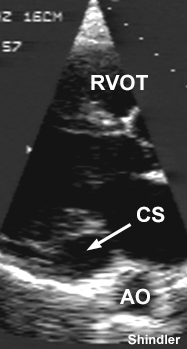
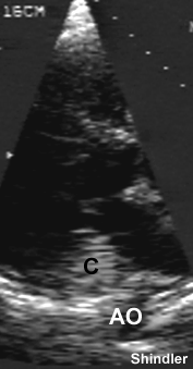
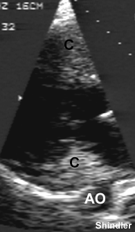
A dilated coronary sinus can be recognized when performing a standard parastenal view.
Sequential images showing a dilated coronary sinus (CS). It has to be distinguished from the descending aorta (AO). Saline contrast (black letter "C") is injected into a left arm vein. The contrast travels from the left arm to a persistent left superior vena cava draining into the coronary sinus. On the echo it appears first in the coronary sinus, then in the right ventricular outflow (RVOT).
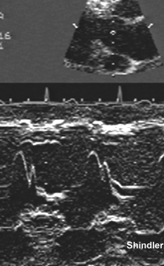 M-mode through the dilated coronary sinus
may confuse by suggesting a large pericardial
effusion.
M-mode through the dilated coronary sinus
may confuse by suggesting a large pericardial
effusion.
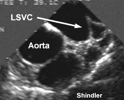
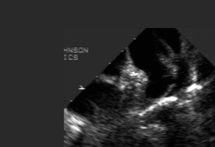 Image loop of a transesophageal study showing
an oval-shaped persistent left superior vena cava
opacified with agitated intravenous saline that was
injected into a left antecubital vein. The left
atrial appendage is to the left. The left upper
pulmonary vein is to the right.
Image loop of a transesophageal study showing
an oval-shaped persistent left superior vena cava
opacified with agitated intravenous saline that was
injected into a left antecubital vein. The left
atrial appendage is to the left. The left upper
pulmonary vein is to the right.
Nsah EN, Moore GW, Hutchins GM. Pathogenesis of persistent left superior vena cava with a coronary sinus connection.
Pediatr Pathol 1991 Mar-Apr;11(2):261-9
The basis for persistence of the left superior vena cava (LSVC), usually associated with cardiac malformations, is poorly understood. The authors examined 351 staged, serially sectioned human embryos in the Carnegie Embryological Collection and 1208 specimens with congenital cardiovascular malformations in the Pathology Collection of the Johns Hopkins Hospital. A standardized questionnaire was answered for each embryo and autopsy case and a computer program was employed to tabulate concurrent anatomic features. In the normal embryos a symmetric venous system appeared with the heart tube at Carnegie stage 9; the sinoatrial junction translocated to the right and the relationship of the coronary sinus to the LSVC was established by stage 12. The LSVC was patent through stage 20 and subsequently underwent luminal obliteration by compression between the left atrium and the hilum of the left lung. Among the 1208 hearts with a congenital abnormality, 104 (9%) had a persistent LSVC with a coronary sinus connection. Statistically, significantly more frequent associations were found between persistent LSVC and atrioventricular canal defects, cor triatriatum, and mitral atresia and a significantly less frequent association was observed between persistent LSVC and atrial septal defect or patent foramen ovale as a primary defect. The normally late embryonic obliteration of the LSVC suggests that its persistence would be secondary to reduce cardiac compression or to blood flow redistribution at an early stage, and the malformations associated with persistent LSVC support that view. Identification of a persistent left superior vena cava with coronary sinus connection should suggest an associated malformation, especially atrioventricular canal, cor triatriatum, or mitral atresia.
Back to E-chocardiography Home Page.

The contents and links on this page were last verified on October 2, 2008.