Quadricuspid Aortic Valve
Tudor Vagaonescu, M.D.
Patricia Ambrosio, R.D.C.S.
Paul Haluska, M.D., Ph.D.
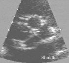
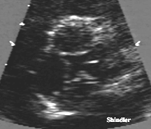
 Transthoracic short axis view showing the diastolic
"X" configuration of the four aortic leaflets.
The right coronary ostium is also visible.
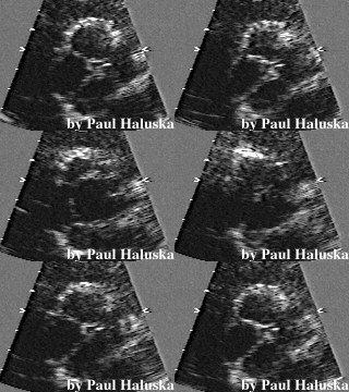
 Partially open quadricuspid aortic valve with a
distinct rectangular orifice.

Incidence
Quadricuspid aortic valve not associated with
truncal abnormalities is rare with a reported
incidence of 0.008% to 0.033%.
(European Heart Journal 1988;9:1269-1270)
In a surgical/pathological series of 225 patients
with pure aortic insufficiency the incidence was 1%.
(Mayo Clin Proc 1984;59:853-841)
Pathology
On pathological descriptions the aortic
valve has four cusps: a posterior cusp, a left
coronary cusp, a right coronary cusp, and an anterior
supernumerary cusp. Raphes join the supernumerary
cusp with the left and right coronary cusps. The raphes
have been described as shallow and chordlike. The
supernumerary cusp can have multiple fenestrations.
(Am J Cardiovascular Pathology 1990;3(2):185) The cusps
can vary in size, thickness, and pliability. (Am J
Cardiovascular Pathology 1990;3(1):91-94)
Associated cardiac abnormalities
-
Displaced coronary ostia
-
Single or accessory coronary ostia
-
Isolated coronary ostium
-
Patent ductus arteriosus
-
Congenitally deficient mitral leaflets
-
Ventricular septal defect
-
Fibromuscular subaortic stenosis
-
One case was associated with non-obstructive
hypertrophic cardiomyopathy. (Heart 1997;78:83-87)
-
Aortic insufficiency is the predominant valvular
abnormality. (Arch Mal Coeur 1996;89:91-93)
It has been postulated that with cusps
of nearly equal area, transvalvular forces are
equally distributed. In contrast, a small accessory
cusp would result in unequal distribution of stress
and consequently may predispose to aortic regurgitation.
(Am J Cardiol 1990;65:937-938) Periodic echocardiographic
evaluations for insufficiency as well as endocarditis
prophylaxis are advisable. (The Journal of Heart Valve Disease
1998;7:515-517)
Embryology
The semilunar valves are derived from mesenchymal
swellings in the aortic and pulmonary
trunk after the truncus arteriosus has been partitioned. It is
in the early stages of truncal separation that four subendothelial
buds appear instead of three. The presence of a corpus arnatii
on all four cusps indicates that the valve resulted from abnormal
embryogenesis. (Am J Cardiol 1991;67:323-324)
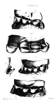
Excess in the number of the semilunar valves.
From Peacock's 1858 book on Malformations of the
Human Heart.
 Fig. 1. Four valves at the orifice of the pulmonary
artery, the excess being apparently produced by the
division of one of the valves at 'a'.
The two segments so produced are imperfect and are
freely blended together.
From a female 75 years of age. The preparation is numbered B 13,
in the Museum of the Victoria Park Hospital.
Fig. 2. Four valves at the orifice of the pulmonary artery;
the excess consisting in three imperfectly divided segments
and one complete segment. The larger fold at 'a', 'a', is
attached to the side of the vessel by firm bands. From a
man, 45 years of age, who was crushed to death.
Fig. 3. Four valves at the aortic orifice, from a preparation
in St. Thomas's Hospital Museum, numbered beta 105. The excess is
apparently due to the division of one fold at 'a' into two.
The septum between these two segments is very imperfect, being,
as seen in fig. 4, perforated by apertures, or displaying
portions in which the fibrous tissue is wanting.
It is doubtful whether the small body marked b, fig. 3, is an
adhesion between the curtains or a supernumerary valve.
Fig. 5. Five valves at the orifice of the pulmonary artery,
from a preparation marked B 12 in Museum of the Victoria Park
Hospital, removed from a child aged four and a half years.
The excess is apparently due to the division of two curtains
at 'a' and 6. The supernumerary segments and those adjacent to
them are imperfect.
Back to E-chocardiography Home Page.
e-mail:shindler@umdnj.edu
The contents and links on this page were last verified on
September 12, 2002.
 Transthoracic short axis view showing the diastolic
"X" configuration of the four aortic leaflets.
 Transthoracic short axis view showing the diastolic
"X" configuration of the four aortic leaflets.
 Partially open quadricuspid aortic valve with a
distinct rectangular orifice.

 Fig. 1. Four valves at the orifice of the pulmonary
artery, the excess being apparently produced by the
division of one of the valves at 'a'.
The two segments so produced are imperfect and are
freely blended together.
From a female 75 years of age. The preparation is numbered B 13,
in the Museum of the Victoria Park Hospital.