 Transesophageal echo showing a persistent left
superior vena cava draining into the left atrium
between the left atrial appendage and the left
upper pulmonary vein. Saline contrast injection
into the left arm confirmed the diagnosis.
Transesophageal echo showing a persistent left
superior vena cava draining into the left atrium
between the left atrial appendage and the left
upper pulmonary vein. Saline contrast injection
into the left arm confirmed the diagnosis.
Raghib et al described eight cases in Circulation 1965;31:906-18 with the following findings:
In this paper the atrial septal defect was considered a true defect of a specific type and associated with absence of the coronary sinus.
The senior author was Dr. Jesse Edwards who had earlier "considered the atrial septal defect of this condition not a true defect of the atrial septum. Rather it was considered that the anterior wall of the coronary sinus was deficient and therefore allowed an interatrial communication." (Gould SE editor, Pathology of the Heart. Second Edition 1960 p. 275)
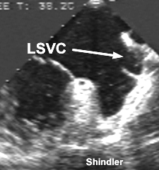
Transesophageal echo showing a persistent left
superior vena cava draining into the left atrium
between the left atrial appendage and the left
upper pulmonary vein. Saline contrast injection
into the left arm confirmed the diagnosis.
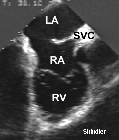 Atrial septal defect in the same patient. A right
superior vena cava was present (SVC) but the
innominate vein was hypoplastic in this patient.
Atrial septal defect in the same patient. A right
superior vena cava was present (SVC) but the
innominate vein was hypoplastic in this patient.
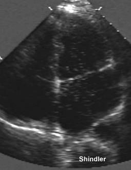 Transthoracic image showing saline contrast that
was injected in the left arm and appeared first
in the left atrium then in the right atrium.
Transthoracic image showing saline contrast that
was injected in the left arm and appeared first
in the left atrium then in the right atrium.
Chen MC, Hung JS, Chang KC, Lo PH, Chen YC, Fu M. Partially unroofed coronary sinus and persistent left superior vena cava: intracardiac echocardiographic observation.
J Ultrasound Med 1996 Dec;15(12):875-9
Unroofed coronary sinus, a rare congenital anomaly first described by Raghib and colleagues in 1965, is a result of an embryologic error involving imperfect or complete failure of development of the left atriovenous fold, which is manifested as a focal (fenestration or partial unroofing of the coronary sinus) or complete absence of the coronary sinus septum. Before the era of echocardiography, precise diagnosis of this anomaly was possible only during surgical procedure or at autopsy. Since the advent of the echocardiography, several studies have reported the usefulness of two-dimensional transthoracic and transesophageal echocardiography in the diagnosis of unroofed coronary sinus. The authors describe the intracardiac echocardiographic delineation of partially unroofed coronary sinus and persistent left superior vena cava in a patient with atrioventricular nodal reentrant tachycardia. Incidental finding of the dilated coronary sinus during radiofrequency ablation of the tachycardia led to the diagnosis of this unusual anomaly.
Okumori M, Hyuga M, Ogata S, Akamatsu T, Otomi S, Ota S. Raghib's syndrome: a report of two cases.
Jpn J Surg 1982;12(5):356-61
The authors treated two patients with a rare developmental complex. The persistent left superior vena cava draining into the left atrium (PLSVC into LA) was associated with an absent coronary sinus and an atrial septal defect. Ligation of PLSVC and patch-repair of the atrial septal defect were successfully performed in one stage. The atrial septal defect was located in the upper and posterior aspect of the interatrial septum and appeared to be an unique type of atrial septal defect. In the other patient, additional multiple cardiac defects were associated with this syndrome, including ventricular septal defect, pulmonary stenosis, tricuspid insufficiency, and complete transposition of the great arteries. Palliative Blalock procedure was used for this patient. The PLSVC into LA was discovered accidentally in both cases during heart catheterization and it was clearly demonstrated by venography. For a preoperative recognition of PLSVC, computed tomograms of the heart are of great assistance. Surgical correction of the persistent superior vena cava was emphasized for treatment of this syndrome.
Fujimura Y, Katoh T, Koizumi S. Raghib's syndrome associated with cor triatriatum - a rare surgical case report.
Nippon Kyobu Geka Gakkai Zasshi 1989 Nov;37(11):2417-21
A 27-year-old male with a history of congenital heart disease was admitted for pre-operative evaluation of a cardiac malformation. Echocardiography and cardiac catheterization revealed an incomplete endocardial cushion defect with a persistent left superior vena cava which drained into the left atrium but echocardiographic evidence of an abnormal intra-atrial septum was not found. The patient was placed on cardiopulmonary bypass and prepared for the surgical correction of his primary cardiac lesion. Intracardiac examination during reconstruction of Raghib's syndrome also revealed the presence of cor triatriatum. Raghib's syndrome is characterized by the combination of abnormal drainage from the left superior vena cava into the left atrium, the presence of an atrial septal defect and the absence of a coronary sinus. To prevent secondary complications such as a brain abscess, the authors redirected blood flow from the left superior vena cava to the right atrium utilizing a trimmed woven dacron vascular graft that was placed in the intra-atrial position. Mitral valvoplasty, excision of the obstructing diaphragm and atrial septation were also performed successfully. Although the literature has described the surgical repair of Raghib's syndrome, its correction in combination with cor triatriatum is considered to be extremely rare.
Honnekeri ST, Tendolkar AG, Lokhandwala YY. Double-orifice mitral and tricuspid valves in association with the Raghib complex.
Ann Thorac Surg 1993 Apr;55(4):1001-2
A 19-year-old woman presented clinically with a left to right pretricuspid shunt. Echocardiography revealed a large ostium primum defect and a double-orifice mitral valve. Operative findings revealed additionally a double-orifice tricuspid valve and a left superior vena cava draining to the roof of the left atrium. The atrioventricular valves were competent. The primum defect was patched and the left superior vena cava was rerouted.
Back to E-chocardiography Home Page.
e-mail:shindler@umdnj.edu
The contents and links on this page were last verified on October 11, 2001.