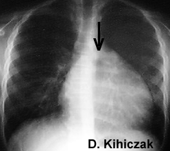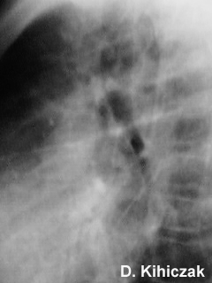


Radiographs of a 12 year old boy with history of rheumatic fever demonstrate cardiomegaly. The lateral view demonstrates the "walking man sign" - posterior displacement of the left main bronchus relative to the right. This is due to the enlarged left atrium, resembling a person walking in mid stride.
Aust N Z J Med. 1994 Oct;24(5):530-5
Doppler echocardiography and the early diagnosis of carditis in acute rheumatic fever.
Abernethy M, Bass N, Sharpe N, Grant C, Neutze J, Clarkson P, Greaves S, Lennon D, Snow S, Whalley G.
Department of Medicine, University of Auckland School of Medicine, New Zealand.
BACKGROUND: The incidence of acute rheumatic fever in New Zealand remains relatively high. Reliable early diagnosis of carditis is difficult and important in management. AIM: To determine if Doppler echocardiography contributed to the early diagnosis of carditis in acute rheumatic fever. METHODS: Forty-seven patients admitted to hospital with suspected acute rheumatic fever and 19 control patients, with a febrile illness due to a documented non-cardiac bacterial infection, were assessed two days and two weeks following admission. Presence or absence of clinical carditis was determined by a cardiologist unaware of the suspected diagnosis, from clinical examination, chest radiograph, electrocardiogram (ECG) and two dimensional echocardiogram. Doppler echocardiography was then performed and interpreted by a second cardiologist unaware of the diagnosis. After completion of the study the Jones criteria were applied, to categorise the patients with suspected acute rheumatic fever into four groups for the final diagnosis: no acute rheumatic fever, possible acute rheumatic fever, definite acute rheumatic fever without carditis, and definite acute rheumatic fever with carditis. RESULTS: In 19 patients with a final diagnosis of acute rheumatic fever and carditis at the baseline assessment carditis was detected by clinical assessment in 15 patients, compared with 19 patients with evidence of significant valve regurgitation by Doppler echocardiography. Following the two week assessment, all 19 patients had both clinical and Doppler evidence of carditis. Five patients with a final clinical diagnosis of possible acute rheumatic fever or definite acute rheumatic fever without carditis, had a Doppler abnormality detected. There was no clinical or Doppler abnormality in the febrile controls. CONCLUSIONS: Doppler echocardiography is more sensitive than clinical assessment in the detection of carditis in acute rheumatic fever, and can contribute to earlier diagnosis.
Back to E-chocardiography Home Page.
e-mail:shindler@umdnj.edu The images and links on this page were last verified on February 24, 2005.