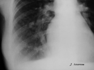

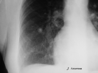
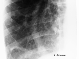
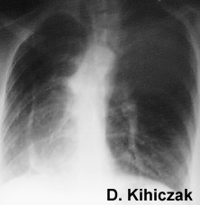
Five patients with scimitar syndrome (three boys, two girls) with a mean age of 3.4 years (range 6 months to 11 years) were studied with two-dimensional and Doppler echocardiography. Four-chamber apical and parasternal short- and long-axis views were obtained, in addition to subcostal views. The anomalous drainage was seen entering the inferior vena cava below the diaphragm in three patients, and in two cases the inferior vena cava was considered normal (both patients had supradiaphragmatic drainage). The anomalous flow velocity pattern was monophasic without reverse flow at atrial contraction. The peak velocity ranged from 0.6 to 1.0 m/s (mean 0.8 m/s). In one case an ostium secundum atrial septal defect was detected. It is concluded that two-dimensional and Doppler echocardiography permit demonstration of the anomalous drainage, analyze the anomalous flow velocity pattern, and detect other associated cardiac abnormalities.
Congenital abnormalities of the tracheobronchial tree are rare causes of recurrent chest infections in childhood. A case is described which shows some typical features of horseshoe lung. More detailed imaging revealed complete separation of the right and left pleural cavities and the malformation represents part of the sequestration spectrum. The case emphasises the need for careful evaluation of the pulmonary arteries, veins, bronchi and oesophagus, particularly prior to surgery.
The authors report 4 cases of the scimitar syndrome with pulmonary hypertension by stenosis of an abnormally draining right pulmonary vein and they also review the literature. All cases were symptomatic from infancy. The diagnosis was confirmed by catheterisation which showed a significant pressure gradient between the right pulmonary vein and the inferior vena cava, and by angiography which demonstrated the stenosis. None of the treatments proposed (interventional catheterisation with dilatation and eventual implantation of a stent, surgery with treatment of the stenosis and reimplantation of the right pulmonary vein in the left atrium, or pneumonectomy) were satisfactory. However, it is possible that earlier treatment could be effective as changes in the pulmonary vascular bed seem to occur very early in these patients.
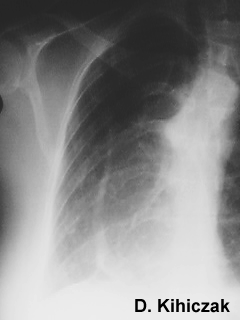
Scimitar syndrome is relatively uncommon syndrome. A 14-year-old female referred to our hospital for abnormal shadow of chest fluoroscopic examination. She had no symptom except cough due to bronchial asthma. Chest X-ray film showed Scimitar sign. Cardiac catheterization revealed 30% left to right shunt without atrial septal defect. Pulmonary angiography showed anomalous pulmonary vein was drained to inferior vena cava and left atrium. Surgical correction was attempted monitoring the pulmonary arterial pressure. Anomalous pulmonary vein and systemic arteries were ligated and divided at the level of diaphragm. Postoperative course was smooth. The authors found 4 similar operated cases previously reported in the world.
The classic definition of the scimitar syndrome is a triad of hypoplasia of the right lung with anomalous venous drainage and a systemic arterial supply of a variable degree. We report a case in which a scimitar-shaped anomalous vein was observed on the plain chest radiograph, but subsequently a pulmonary angiogram showed that it drained normally into the left atrium.
In 121 adults, the value of transthoracic and transesophageal color Doppler echocardiography for detection of different types of atrial septal defect (ASD) or of partial anomalous pulmonary venous return was analyzed. The 121 patients had a total of 129 defects with left-to-right atrial shunting (including eight patients with two types of defects). All of six cases with primum-type ASD were diagnosed correctly by both echocardiographic methods. Ninety-seven patients showed a secundum-type ASD during transesophageal echocardiography: by transthoracic echocardiography, only eight (20%) of the 40 small defects (diameter < 5 mm) were detected as compared with 15 (83%) of the 18 defects with a diameter of 5 to 10 mm and all 39 defects with a diameter > 10 mm. A sinus venosus--type ASD was evident by transesophageal echocardiography in 11 patients, of which only one (9%) was demonstrated by the transthoracic approach. Partial anomalous pulmonary venous return was seen by transesophageal echocardiography in 13 patients but missed in two other patients in whom anomalous pulmonary venous return was subsequently identified by surgery (both with anomalous return of the upper right pulmonary vein into the superior vena cava). By use of the transthoracic technique, partial anomalous venous return was detected in only two cases, both of which had "scimitar syndrome." Compared with transthoracic echocardiography, the transesophageal approach is clearly superior in the detection of small secundum-type ASD, sinus venosus--type ASD, and partial anomalous pulmonary venous return.
Horseshoe lung is an uncommon congenital malformation in which the bases of the right and the left lungs are fused to each other by a narrow isthmus posterior to the cardiac apex. So far 22 cases have been described: most of these were associated with right lung hypoplasia and the scimitar syndrome. A horseshoe lung anomaly with left lung hypoplasia is described.
One hundred twenty-two cases of the adult form of the scimitar syndrome were collected from different cardiologic centers. The clinical, radiographic and hemodynamic findings are described. The scimitar syndrome is defined as an anomalous right pulmonary venous drainage, partial or complete, to the inferior vena cava. Additional characteristics of this syndrome such as hypoplasia and abnormalities of the vascular supply to the right lung, dextrocardia and abnormalities of the bronchial segmentation are common; bronchiectases are rare. The left to right shunt was less than 50% in 100 of the 122 patients. The pulmonary arterial pressures were normal in 94 patients and slightly elevated in 28. A follow-up study of these patients showed that, without surgical correction, they lead a normal life. An awareness of this syndrome may avoid unnecessary invasive diagnostic procedures and surgical treatment for most patients.
The clinical spectrum of the scimitar syndrome ranges from severely ill infants to asymptomatic adults. The true incidence of the disorder is unknown because the syndrome may remain undetected in asymptomatic patients until a chest roentgenogram is obtained. We have presented the contrasting clinical experiences of two adult women, one with few symptoms and a benign course, and the other with exacerbation of her asthma from recurrent upper respiratory tract infections originating in the lower lobe of her right lung. Improvement resulted from surgical resection of this congenitally abnormal, bronchiectatic segment of lung.
The scimitar syndrome, first described by Chassinat in 1836, consists aessentially of an anomalous pulmonary vein draining whole or part of the right lung into the inferior vena cava. Associated anomalies are frequent, such as hypoplasia of the right lung, dextrocardia, malformations of the right pulmonary artery and bronchial tree, and abnormal arterial supply of the right lung (the so-called sequestration). This article describes a scimitar syndrome associated with stenosis of the inferior vena cava, whose initial diagnosis was made by two-dimensional echocardiographic Doppler color flow mapping. This is the first description of such an unusual association.
A case of the scimitar sign due to an anomaly of the right sided pulmonary vein with normal drainage into the left atrium was associated with an azygos continuation of the inferior vena cava. Digital subtraction angiography allows the identification of these rare congenital vascular malformations.
A case of scimitar syndrome with pulmonary sequestration is reported. Anomalous pulmonary venous return from the right lung to the infradiaphragmatic vena cava inferior was diagnosed by pulmonary angiogram and sequestration of the right lower lobe was confirmed by aortogram. Venous return from the sequestrated lung was partly into the vena cava inferior and partly into the left atrium. Successful repair was achieved by resection of the sequestrated lobe and direct reimplantation of the scimitar vein into the left atrium. Accurate preoperative diagnosis and intraoperative evaluation of the anatomy is mandatory to correct this rare anomaly.
In the classic scimitar syndrome, a pulmonary vein draining all or part of the right lung enters the inferior vena cava. A variant is described with the same roentgenographic appearance, but with drainage of the anomalous pulmonary vein into both the inferior vena cava and the left atrium; the atrial septum was intact. This case, together with six others reported elsewhere, reminds us that the scimitar sign has both false positives and false negatives. Therefore, the diagnosis of scimitar syndrome cannot be made with certainty from a plain x-ray film.
A "horseshoe" lung is a rare congenital anomaly in which the posterobasal segments of the right and left lungs are fused behind the pericardial reflection at the cardiac apex. Most patients with such pulmonary fusion share many of those cardiovascular anomalies typical of the "scimitar" or hypogenetic right lung syndrome. The clinical diagnosis of horseshoe lung can be made by pulmonary angiography, which shows a branch of the right pulmonary artery originating from its proximal but inferior aspect and coursing into the left hemithorax. Thus, there are two conditions in which an artery from the right lung supplies part or all of the left lung: errant right pulmonary artery in the horseshoe lung deformity and so-called left pulmonary artery sling.
A review of the roentgenograms and clinical records of 33 children with primary congenital underdevelopment of one lung showed that 9 patients had simple pulmonary hypoplasia, 8 had anomalous venous return to the right atrium or the inferior vena cava (scimitar syndrome), 7 had an absence of the ipsilateral pulmonary artery, 7 had an accessory diaphragm, and 2 had a pulmonary sequestration adjacent to a small diaphragmatic hernia.
Report on a 28-year-old female patient with classic "scimitar syndrome" (dextroposition of the heart, hypoplasia of the right lung and pulmonary veins opening into the inferior vena cava). The condition was diagnosed on the basis of chest X-ray findings in which the most striking feature was the scimitar-like course of the right pulmonary veins which gives the syndrome its name. The diagnosis was confirmed and the shunt volume determined by cardiac catheterization. Since in this case there were no additional deformities such as atrial septum defect, ventricular septum defect or patent ductus botalli and the shunt volume was small, surgical correction was not required.
Back to E-chocardiography Home Page.

The contents and links onthis page were last verified on October 22, 2012
by Dr. Olga Shindler