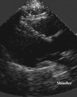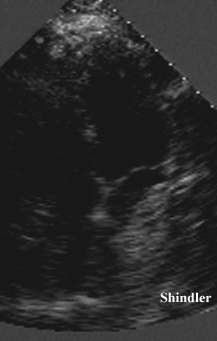
Scoliosis is associated with mitral valve prolapse. Echocardiographic windows may not be in their customary location. The spine may impinge on the left atrium as shown below.


Clinical characteristics of 60 (41 males, 19 females) patients with echocardiographically proven mitral valve prolapse were analysed, with special interest in the associated thoracic skeletal abnormalities. There was a male preponderance (2.2:1) and 91.7% of patients were symptomatic--atypical chest pain, palpitations, exertional dyspnoea and easy fatiguability being the major symptoms. Sixty seven percent had an asthenic body habitus, and 55% had high-arched palate. Thoracic scoliosis (55%), straight back syndrome (50%), flat chest (46.7%), and pectus excavatum (20%) were seen in association with the condition, with 81.7% having any one or combination of these features. Lateral chest radiography showed pancaking of heart shadow in 48.3%. Isolated non-ejection systolic click(s) was the major cardiac auscultatory finding (61.7%), while 60% showed pansystolic prolapse on echocardiography. Electrocardiographic ST-T-U changes in the inferior and/or lateral chest leads were seen in 46.7%, while 16.7% had cardiac arrhythmias. None had infective endocarditis, heart failure or cerebral embolic events. The findings corroborate the view that thoracic skeletal anomalies may be regarded as non-auscultatory features of this syndrome.
Dhuper S, Ehlers KH, Fatica NS, Myridakis DJ, Klein AA, Friedman DM, Levine DB. Incidence and risk factors for mitral valve prolapse in severe adolescent idiopathic scoliosis. Pediatr Cardiol 1997 Nov-Dec;18(6):425-8
Mitral valve prolapse (MVP) is known to be associated with thoracic skeletal anomalies. To determine the incidence and risk factors for mitral valve prolapse in the adolescent population with severe idiopathic scoliosis (IS), a prospective follow-up study on 139 adolescent patients with IS from the Pediatric Orthopedic Service was undertaken. Data collected included age, sex, medical and family history, physical exam, electrocardiogram and echocardiogram, spinal x-rays, and pulmonary function tests. MVP w as detected by echocardiogram in 13.6% (19/139) of patients with IS as compared with 3.2% in 154 age- and weight-matched controls (p < 0.006). All patients with MVP were asymptomatic and a systolic click or murmur was detected on the single preoperative e xam only in 37% (7/19) of them. Patients with MVP and IS weighed less (45.1 +/- 2.0 vs 51.8 +/- 0.1 kg, p < 0.002) as compared with those IS patients without MVP. The electrocardiogram was abnormal in 21% (4/19) of patients with MVP as compared with only 1.6% (2/120) of patients with IS but no MVP. The two groups did not differ with respect to age at diagnosis, severity of scoliosis, positive family history of scoliosis, or the presence of restrictive lung disease. Though IS was more prevalent in females (79%), the presence of MVP was not related to gender. MVP was persistent in 10 of the 19 patients reevaluated by echocardiogram 2-4 years after spinal surgery. We conclude that MVP is four times more common in patients with severe IS than in the normal ad olescent population, and is associated with a lower body weight in IS patients with MVP than in IS patients without MVP. The persistent nature of MVP, even after corrective spinal surgery, may be related to factors other than geometric changes of the hear t caused by abnormal thoracic curvature.
Thirty-six children and adolescents with early stages of idiopathic scoliosis underwent evaluation by echocardiography and pulmonary function testing. Mildly increased pulmonary vascular resistance was inferred from an elevated ratio of right preejection period to right ventricular ejection time, an increased right ventricular dimension, and a decreased left ventricular dimension. Since neither decreased arterial oxygen saturation nor increased end-tidal expired carbon dioxide partial pressure was seen, desaturation and hypoventilation should not account for these abnormalities. Pulmonary function parameters showed no distinct patterns of abnormality. Even though the patients were divided into two groups by severity of spinal curvature, the cardiopulmonary measures did not correlate with thoracic deformity. Billowing of the mitral leaflets, termed mitral valve prolapse, was demonstrated in 25% of the subjects. Our findings suggest that cardiopulmonary and thoracic changes in idiopathic scoliosis may develop in parallel and may be expressions of a common collagen defect. However, study of sleep and exercise arterial saturation may be required to rule out intermittent hypoxemia as a precipitating factor of cor pulmonale in scoliosis.
Seventy-four patients with adolescent scoliosis underwent cardiac examination and M-mode echocardiography to detect the presence of mitral valve prolapse (MVP). Twenty-one (28%) had echocardiographic evidence of MVP, whereas 18 had auscultatory findings of a nonejection click or late systolic murmur. A subset of 41 patients had a family history of scoliosis and 37% had MVP. The incidence of MVP increased to 41% when a first degree relative, such as a sibling, parent, or offspring, had scoliosis. Thirty-six patients with scoliosis had additional thoracic hypokyphosis (straight back) and 13 (36%) had MVP. The incidence of MVP was 48% when the scoliosis and hypokyphosis were hereditary and increased to 53% when a familial history of skeletal abnormality was present. This study indicates a high incidence of MVP in patients with scoliosis and hypokyphosis, especially when the cardiac and skeletal systems may be affected by a generalized soft-tissue defect.
Idiopathic prolapse of the mitral valve is a common disorder, but many cases are clinically subtle. Thoracic skeletal abnormalities, reported recently to accompany the syndrome, may serve as an easily identifiable clinical indicator. The prevalence of these abnormalities was defined in 24 patients with proved prolapse of the mitral valve. The valvular syndrome was defined clinically, by echocardiography and, in seven cases, by left ventricular angiography. The skeletal deformities were defined clinically and radiographically. Pectus excavatum was present in 62 percent of the patients, "straight back" in 17 percent and severe scoliosis in 8 percent. Eighteen of the 24 patients (75 percent) had a definite thoracic skeletal deformity. The association of idiopathic prolapse of the mitral valve with these skeletal deformities may represent a forme fruste of Marfan's syndrome. Patients with "straight back" and pectus excavatum should be examined clinically and perhaps by echocardiography to exclude idiopathic prolapse of the mitral valve; when murmurs are present, a diagnosis of "pseudoheart disease" should not be made before mitral valve prolapse has been excluded.
Department of Anaesthesiology, Hospital Universitario Vall d'Hebron, Passeig Vall d'Hebron, 119-129, 08035, Barcelona, Spain.
e-mail:26750mjc@comb.es
The prevalence of asymptomatic cardiac valve anomalies was determined in 82 patients (69 females and 13 males) diagnosed as having idiopathic scoliosis and scheduled for corrective surgery (mean age at surgery 16.3 years). The preoperative study in each p atient included echocardiography and ultrasound Doppler. Twenty-three valvular anomalies were found in 20 patients (24.4%). The most frequent was mitral valve prolapse. The occurrence of valvular anomalies did not correlate with sex, curve magnitude, or a ge at diagnosis. Eighteen patients presented a total of 20 comorbid conditions: positive family history of scoliosis (five cases), isthmic spondylolisthesis (five cases), nervous anorexia (two cases), hereditary exostosis, cystic fibrosis, ureteral stenos is, mammary hypoplasia, slipped capital femoral epiphysis, psoriasis, celiac disease, and lactose intolerance. A significant relationship was found between valvular anomalies and comorbidity. Valvular anomalies were detected in 11 out of 64 patients (17.2 %) with no comorbidity and in nine out of 18 patients (50%) with a comorbid condition (Chi-square 8.2, p = 0.004). In this latter group of patients, routine echocardiographic study seems advisable in the preoperative evaluation.
Back to E-chocardiography Home Page.

The contents and links on this page were last verified on October 11, 2002.