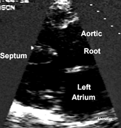
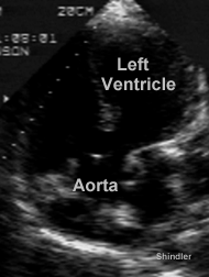
Tetralogy of Fallot described in a case report in the first issue of the New England Journal of Medicine. In a letter from John S. Dorsey, M.D. Adjunct Professor of Surgery in the University of Pennsylvania
S. R. when born, was for a considerable time supposed to be dead - did not cry, or evince any living actions. The lungs were artificially inflated for several minutes, and life at length appeared, but very feebly. A livid countenance, with frequent syncope took place. With great maternal care the infant was kept alive, and as she grew became remarkably sprightly and active. When two years old was unusually intelligent and fond of exercise. As she advanced in age, her fondness for violent exercise in playing often exposed her to danger, as these efforts never failed to produce syncope and a kind of convulsion. Laughing, crying, or any emotion of mind, also brought on the syncope, from which, after falling into a horizontal position, she generally soon recovered. Her countenance, at all times blueish and livid, was in these fits extremely so. Her nails were always of the colour of litmus, or perhaps a little nearer to violet. She had the usual diseases of children, the vaccine-chicken pox, scarlatina, whooping cough, measles from all which she recovered as rapidly as is usual. The peculiarities of these children in whom the foramen ovale of the heart remains open, all appeared in this little girl, and need not be more minutely described. After death the thorax was examined. It was of an unusual shape, being more cylindrical than common, and the lungs having less the form of a cloven hoof, when inflated, than they unusually assume. The heart was very small. In place of a right auricle was observed a small appendage like the edge of that portion of the heart, not capable of containing more than one fourth its usual contents. The right ventricle was as firm in texture as the left, and the quantity of muscular substance about equal in both ventricles. But the most singular circumstance was in the distribution of the great arteries. The pulmonary was extremely small. The aorta of unusual size, and communicating with both ventricles.
Back to E-chocardiography Home Page.
e-mail:shindler@umdnj.edu The contents and links on this page were last checked on December 18, 2002.