Danbauchi S S, MBBS, FWACP
Consultant Cardiologist/Senior Lecturer
Cardiology Unit, Department of Medicine
Ahmadu Bello University Teaching Hospital Zaria
Anumah F E, MBBS, FMCP
Consultant Endocrinologist/Lecturer I
Endocrine Unit, Department of Medicine
Ahmadu Bello University Teaching Hospital Zaria
Alhassan M A, MBBS, FMCP
Consultant Cardiologist/Lecturer I
Cardiology Unit, Department of Medicine
Ahmadu Bello University Teaching Hospital Zaria
Oyati I A, MBBS
Senior Registrar/Lecturer II
Cardiology Unit, Department of Medicine
Ahmadu Bello University Teaching Hospital Zaria.
Professor Isah H S, BSc, MSc, PhD
H O D, Chemical Pathology
Ahmadu Bello University Teaching Hospital Zaria
Onyemelukwe G C, FMCP, FWACP, FICS, MON
Head, Immunology Unit Department of Medicine
Ahmadu Bello University Teaching Hospital Zaria
Tiam E A
Senior Medical Laboratory Scientist,
Department of Chemical Pathology
Ahmadu Bello University Hospital, Zaria.
Correspondence to
Danbauchi S S
Cardiology Unit, Department of Medicine
Ahmadu Bello University Teaching Hospital Zaria, Nigeria.
e-mail
danbauchi@skannet.com
SUMMARY
Eleven patients out of 132 with Graves' disease presented with symptoms and signs of thyrocardiac disease from May 1999 to December 2002. There were nine females and 2 males with a mean age of 38.0 +/- 7.8. They all had normal or low BMI. There were eight patients with systolic hypertension and three with atrial fibrillation. Eight patients had normal ejection fraction (high output cardiac failure), three had cardiomegaly, and three had diastolic dysfunction. Treatment with carbimazole improved symptoms, thyroid function tests and echocardiographic features, except in one of the patients with cardiac dilatation who remained in heart failure.
INTRODUCTION
The combination of hypervolemic burden, reduced myocardial contractile reserve, and tachyarrhythmias can result in cardiac failure. (1) It has been suggested that thyrotoxicosis causes a reversible cardiomyopathy although no destructive myocardial lesions have been identified. (2)
We describe the clinical and echocardiographic findings in our subjects with thyrocardiac disease.
METHODS
Patients who were diagnosed with hyperthyroidism (thyrotoxicosis) and in addition presented with symptoms and signs of cardiac decompensation such as shortness of breath, paroxysmal nocturnal dyspnoea, cough, palpitations, leg and abdominal swelling, S3 or S4 gallop, and crepitations were recruited to the study which lasted from June 1999 to May 2002. Hyperthyroidism was established by measuring thyroid function and cardiac failure was diagnosed using clinical signs as defined by Framingham criteria. (3) Hypertension was defined by the current WHO criteria for systolic, diastolic or both systolic and diastolic cut off points. (4) All the patients had Graves' disease.
Basic investigations were conducted in addition to standard 12 lead electrocardiography (ECG), chest radiograph (PA view) and echocardiography. The echocardiographic parameters recorded included aortic root diameter (AR), left ventricular dimensions in diastole and systole, end diastolic volume (EDV) and end systolic volume (ESV), ejection fraction (EF) and fractional shortening (FS). Doppler flow studies at the mitral valve inflow area were conducted to evaluate E and A wave velocity. The data were reported as percentages for non-quantitative parameters and means +/- standard deviation (SD).
RESULTS
From May 1999 to December 2002 a total of 728 requests for thyroid function tests were made to the Chemical Pathology Laboratory at Ahmadu Bello University Teaching Hospital, Zaria. One hundred and thirty two (18.2%) were found to have hyperthyroidism (levels above the reference values for Zaria). Eleven (8.3%) of 122 patients also had clinical symptoms and signs of cardiac failure. They were made up of nine females and two males, with a ratio of 4.5 to 1. The mean age and BMI were 38.0 +/- 7.8 (SD) years and 23.5 +/- 1.8 Kg/m2 respectively. Nine patients had systolic hypertension and two had diastolic hypertension based on WHO criteria.
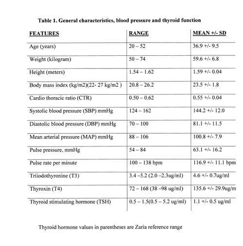
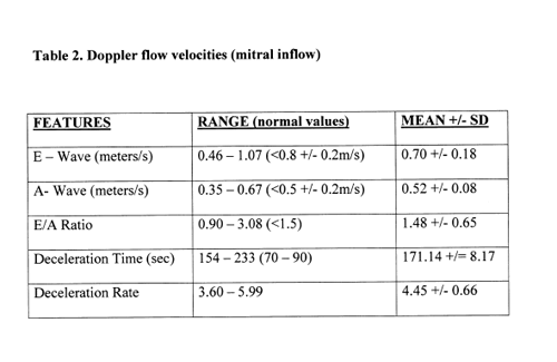
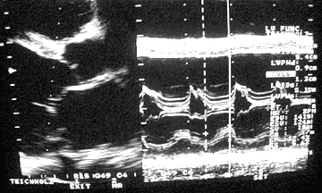
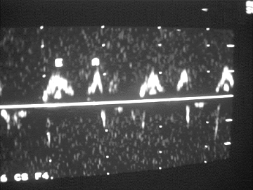
Thyroid function tests showed means of T3, T4 and TSH of 4.6 +/- 0.68 (2.0 –2.3 ug/ml, reference values), 135.56 +/- 29.95 (38 – 98 ug/ml, reference values) and 1.1 +/- 0.5 (05 – 5.2 ug/ml, reference values) respectively (table 1). The mean cardio-thoracic ratio was 0.55 +/- 0.04 while four patients had definite cardiomegaly (CTR > 0.55). Three and two patients had atrial fibrillation and left ventricular hypertrophy respectively on ECG. The mean of left atrial diameter was 3.12 +/- 0.41 and the mean aortic root diameter was of 2.72 +/- 0.38 cm. The EDV ranged from 91 – 156 mls with a mean of 95.50 +/- 8.17 (normal values < 150 mls) and ESV ranged from 27 – 74 mls with a mean of 37.11 +/- 15.61. The ejection fraction ranged from 28.6 – 73.0 % with a mean of 58.2 +/- 17.1 (normal values > 50 %) and the fractional shortening ranged from 10.6 – 38.9 % with a mean of 27.5 +/- 9.2 (normal values > 18%). Three patients had depressed EF and FS (EF < 50% and FS < 18%) suggesting thyrocardiac cardiomyopathy (Fig 1) in addition they had cardiomegaly on chest radiography. Five patients had septal hypertrophy (septal thickness > 1.2 cm). In addition they had a longer duration of symptoms and cardiomegaly on chest radiography. Doppler revealed three patients with diastolic dysfunction, two in the depressed and one in the normal EF subgroups (reverse E/A ratio and prolonged deceleration time)(Fig 2). Three patients had dilated hearts with depressed EF and FS whereas eight had normal ejection fraction. The patients were started on carbimazole, four patients did remarkably well, except for the one patient with cardiomyopathy who still had persistent symptoms of cardiac failure, and the EF/FS have not appreciated remarkably. The four patients that had improved symptoms also had normal T3 and T4.


DISCUSSION
An incidence of eleven thyrocardiac patients (8.3%) in three and half years from our centre is similar to the report of congestive cardiac failure in hyperthyroid patients of 6.2% by Wilson et al and higher than the 20 cases of thyrocardiac patients from the Congo in a period of nine years. (5,6) The male/female ratio in our subjects is in favour of females similar to ratios in hyperthyroidism. The mean age of our subjects was lower than the report by Okesieme though their report was not restricted to thyrocardiac disease. (7) Our incidence is similar to Famuyiwa’s report. (8)
Thyrocardiac disease is said to be more common in individuals above the age of sixty (9) and probably more common in males than females. Our report showed more females than males and our subjects were mostly below the age of fifty.
There are several diagnostic modalities for the diagnosis of thyrocardiac disease. Kolawole et al have highlighted the importance of chest radiography, electrocardiography and echocardiography. (10) Thyrocardiac cardiomyopathy may present with diastolic or systolic dysfunction. (11) The changes in cardiac function are said to be mediated by T3 regulation of cardiac specific genes and cardiac failure results from poor control of hyperthyroidism. (12) Eight of our patients, though clinically in cardiac failure, had normal ejection fraction and fractional shortening which suggested high output cardiac failure. Three patients had dilated left ventricles with two showing improved symptoms and improved echocardiographic and thyroid function test parameters on treatment. The remaining patient had symptoms suggesting persistent cardiac decompensation.
Ebisawa et al reported four cases of irreversible thyrocardiac cardiomyopathy in adults. (13) Three of our patients had atrial fibrillation. Arrhythmia is known to be the most common presentation of thyrocardiac disease and atrial fibrillation is said to be mostly seen in patients above the age of sixty. (9)
Systolic hypertension has been suggested to be due to the inability of vasculature to accommodate the increase in cardiac output and stroke volume, a mechanism that explains the infrequent presence of diastolic hypertension. (14)
Mitral regurgitation was noted in all our patients similar to the report by Nkoua et al (6) but mitral valve prolapse was not seen. It is reported to increase with advance in age in hyperthyroid patients.
The low mean age of our subjects may explain the paucity of complications like atrial fibrillation, mitral valve prolapse and thrombus in the cardiac chambers.
Figure 1. Parasternal long axis 2D and M Mode echocardiogram of one of the patients with left ventricular dilatation and decreased ejection fraction.

Figure 2. Doppler flow tracing in the mitral inflow area showing reversed E/A ratio indicating diastolic dysfunction.

REFERENCES
Back to E-chocardiography Home Page.

The contents and links on this page were last verified on September 6, 2003.