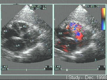
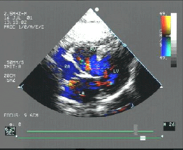
 Three days post rupture - the ventricular septal defect
size is small and the flow is turbulent in nature.
Dr. D. Kannan
S.S.M. Hospital
Kollam - 691001
Kerala, India.
A 52 year male was admitted with acute inferior wall myocardial infarction. The patient had recurrent postinfarct angina and one episode of ventricular fibrillation that required electrical defibrillation. A new systolic murmur was noted after the procedure. Bedside echocardiogram revealed basal ventricular septal rupture.
 Three days post rupture - the ventricular septal defect
size is small and the flow is turbulent in nature.
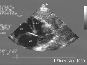 Three weeks post rupture - the defect size has increased
to 2.9 cm. The defect edges are irregular. During this
period the patient had a second episode of ventricular
fibrillation that once again responded to electrical defibrillation.
He remained hypotensive for five days and was diagnosed with
pulmonary embolism (in spite of adequate anticoagulation).
 Three weeks post rupture - color flow shows a laminar flow
pattern with predominant left to right shunt.
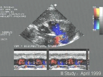 Three months post rupture - color flow reveals a bidirectional
shunt.
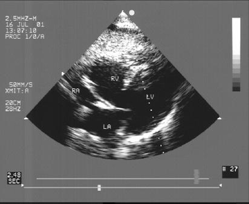 Three years post rupture - the defect size is the same and the
edges are smooth.
 Three years post rupture - color flow reveals bidirectional shunt
with predominant right to left flow.
Back to E-chocardiography Home Page.
e-mail:shindler@umdnj.edu