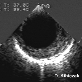
Transesophageal echocardiographic animations of a descending aortic dissection.

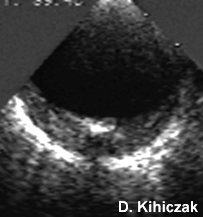
Thrombosed descending aortic dissection with intimal calcification below.
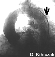
Aortogram showing calcification on the inwardly displaced intima.
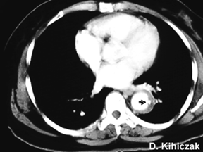
CT (above) showing the same intimal displacement by the thrombosed false lumen. Arrow points to the intimal calcification which appears linear on the longitudinal ultrasound view below.
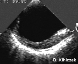
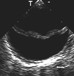
Double lumen in the descending aorta.
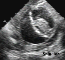
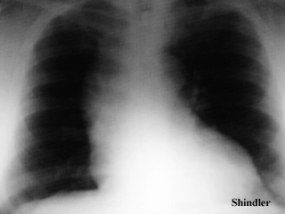
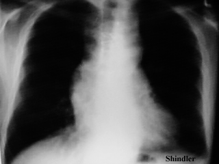
Wide mediastinum on chest x-ray.
Back to E-chocardiography Home Page.

The contents and links on this page were last verified on February 17, 2005.